Coagulation Factor III/Tissue Factor Antibody (TF9-10H10) - BSA Free, Novus Biologicals™
Manufacturer: Fischer Scientific
Select a Size
| Pack Size | SKU | Availability | Price |
|---|---|---|---|
| Each of 1 | NB10065221-Each-of-1 | In Stock | ₹ 49,884.50 |
NB10065221 - Each of 1
In Stock
Quantity
1
Base Price: ₹ 49,884.50
GST (18%): ₹ 8,979.21
Total Price: ₹ 58,863.71
Antigen
Coagulation Factor III/Tissue Factor
Classification
Monoclonal
Concentration
1.0 mg/mL
Dilution
Western Blot 1-10 ug/ml, Flow Cytometry 1:10-1:1000, Immunohistochemistry 1:10-1:500, Immunocytochemistry/Immunofluorescence 1:10-1:500, Immunohistochemistry-Frozen 1:10-1:500
Gene Alias
CD142, CD142 antigen, Coagulation factor III, coagulation factor III (thromboplastin, tissue factor), FLJ17960, TF, TFA, Thromboplastin, tissue factor
Host Species
Mouse
Purification Method
Protein G purified
Regulatory Status
RUO
Primary or Secondary
Primary
Test Specificity
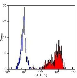
CD142, also known as Tissue Factor, is the membrane receptor for coagulation factors VII and VIIa and is the cell surface initiator of coagulation. It is the major molecule of this type and is criticial for controlling hemostasis, thrombosis and inflammation. NB100-65221 is specific for an epitope within the extracellular domain, epitope locus I. It recognizes both the reduced and native non-reduced human and primate tissue factors. It does not inhibit coagulation or neutralize factor VII binding to CD142.
Content And Storage
Store at 4C short term. Aliquot and store at -20C long term. Avoid freeze-thaw cycles.
Isotype
IgG1
Applications
Western Blot, Flow Cytometry, Immunohistochemistry, Immunocytochemistry, Immunofluorescence, Immunohistochemistry (Frozen)
Clone
TF9-10H10
Conjugate
Unconjugated
Gene Accession No.
P13726
Gene Symbols
F3
Immunogen
Denatured Tissue factor isolated from human brain by the Factor VII affinity method.
Quantity
0.1 mg
Research Discipline
Cancer
Gene ID (Entrez)
2152
Target Species
Human, Primate
Form
Purified
Related Products


Description
- Coagulation Factor III/Tissue Factor Monoclonal specifically detects Coagulation Factor III/Tissue Factor in Human, Primate samples
- It is validated for Western Blot, Flow Cytometry, Immunohistochemistry, Immunocytochemistry/Immunofluorescence, Immunohistochemistry-Frozen.
Compare Similar Items
Show Difference
Antigen: Coagulation Factor III/Tissue Factor
Classification: Monoclonal
Concentration: 1.0 mg/mL
Dilution: Western Blot 1-10 ug/ml, Flow Cytometry 1:10-1:1000, Immunohistochemistry 1:10-1:500, Immunocytochemistry/Immunofluorescence 1:10-1:500, Immunohistochemistry-Frozen 1:10-1:500
Gene Alias: CD142, CD142 antigen, Coagulation factor III, coagulation factor III (thromboplastin, tissue factor), FLJ17960, TF, TFA, Thromboplastin, tissue factor
Host Species: Mouse
Purification Method: Protein G purified
Regulatory Status: RUO
Primary or Secondary: Primary
Test Specificity: CD142, also known as Tissue Factor, is the membrane receptor for coagulation factors VII and VIIa and is the cell surface initiator of coagulation. It is the major molecule of this type and is criticial for controlling hemostasis, thrombosis and inflammation. NB100-65221 is specific for an epitope within the extracellular domain, epitope locus I. It recognizes both the reduced and native non-reduced human and primate tissue factors. It does not inhibit coagulation or neutralize factor VII binding to CD142.
Content And Storage: Store at 4C short term. Aliquot and store at -20C long term. Avoid freeze-thaw cycles.
Isotype: IgG1
Applications: Western Blot, Flow Cytometry, Immunohistochemistry, Immunocytochemistry, Immunofluorescence, Immunohistochemistry (Frozen)
Clone: TF9-10H10
Conjugate: Unconjugated
Gene Accession No.: P13726
Gene Symbols: F3
Immunogen: Denatured Tissue factor isolated from human brain by the Factor VII affinity method.
Quantity: 0.1 mg
Research Discipline: Cancer
Gene ID (Entrez): 2152
Target Species: Human, Primate
Form: Purified

Antigen: CD2
Classification: Monoclonal
Concentration: __
Dilution: Flow Cytometry, Immunocytochemistry/Immunofluorescence, Immunohistochemistry-Paraffin, Immunohistochemistry-Frozen
Gene Alias: CD2 antigen, CD2 antigen (p50), sheep red blood cell receptor, CD2 molecule, Erythrocyte receptor, FLJ46032, LFA-2, LFA-3 receptor, lymphocyte-function antigen-2, Rosette receptor, SRBC, T11, T-cell surface antigen CD2, T-cell surface antigen T11/Leu-5
Host Species: Mouse
Purification Method: Protein G purified
Regulatory Status: RUO
Primary or Secondary: Primary
Test Specificity: NB100-65228 recognizes the rat CD2 cell surface antigen, a 50-54kD glycoprotein expressed by thymocytes and mature T cells. * Whiteland, J.L. et al. (1995). Immunohistochemical Detection of T-Cell Subsets and other Leucocytes in Paraffin-embedded Rat and Mouse Tissues with Monoclonal Antibodies. J. Histochem. Cytochem. 43: 313-320.
Content And Storage: Store at 4C in the dark.
Isotype: IgG2a
Applications: Flow Cytometry, Immunocytochemistry, Immunofluorescence, Immunohistochemistry (Paraffin), Immunohistochemistry (Frozen)
Clone: OX-34
Conjugate: DyLight 488
Gene Accession No.: __
Gene Symbols: CD2
Immunogen: Activated rat T helper cells
Quantity: 0.25 mL
Research Discipline: Adaptive Immunity, Apoptosis, Immunology
Gene ID (Entrez): 914
Target Species: Rat
Form: Purified
Antigen: CD2
Classification: Monoclonal
Concentration: __
Dilution: Immunohistochemistry-Paraffin, Immunohistochemistry-Frozen
Gene Alias: CD2 antigen, CD2 antigen (p50), sheep red blood cell receptor, CD2 molecule, Erythrocyte receptor, FLJ46032, LFA-2, LFA-3 receptor, lymphocyte-function antigen-2, Rosette receptor, SRBC, T11, T-cell surface antigen CD2, T-cell surface antigen T11/Leu-5
Host Species: Mouse
Purification Method: Protein G purified
Regulatory Status: RUO
Primary or Secondary: Primary
Test Specificity: NB100-65228 recognizes the rat CD2 cell surface antigen, a 50-54kD glycoprotein expressed by thymocytes and mature T cells. * Whiteland, J.L. et al. (1995). Immunohistochemical Detection of T-Cell Subsets and other Leucocytes in Paraffin-embedded Rat and Mouse Tissues with Monoclonal Antibodies. J. Histochem. Cytochem. 43: 313-320.
Content And Storage: Store at 4C in the dark.
Isotype: IgG2a
Applications: Immunohistochemistry (Paraffin), Immunohistochemistry (Frozen)
Clone: OX-34
Conjugate: HRP
Gene Accession No.: __
Gene Symbols: CD2
Immunogen: Activated rat T helper cells
Quantity: 0.25 mL
Research Discipline: Adaptive Immunity, Apoptosis, Immunology
Gene ID (Entrez): 914
Target Species: Rat
Form: Purified
