Histone H2AX, p Ser139 Antibody (3F2), Novus Biologicals™
Manufacturer: Novus Biologicals
Select a Size
| Pack Size | SKU | Availability | Price |
|---|---|---|---|
| Each of 1 | NB10074435-Each-of-1 | In Stock | ₹ 54,156.50 |
NB10074435 - Each of 1
In Stock
Quantity
1
Base Price: ₹ 54,156.50
GST (18%): ₹ 9,748.17
Total Price: ₹ 63,904.67
Antigen
Histone H2AX (p Ser139)
Classification
Monoclonal
Concentration
1 mg/mL
Dilution
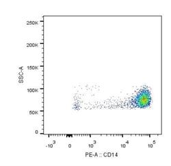
Western Blot 1 ug/ml, Simple Western 10 ug/ml, Flow Cytometry 1 ug 10^6 cells, ELISA 1:100 - 1:2000, Immunohistochemistry 1:10 - 1:500, Immunocytochemistry/Immunofluorescence 2 - 4 ug/ml, Immunohistochemistry-Paraffin 1:10 - 1:500
Gene Alias
H2A.X, H2A/X, H2AFX
Host Species
Mouse
Molecular Weight of Antigen
15 kDa
Quantity
100 μg
Research Discipline
Checkpoint signaling, DNA Double Strand Break Repair, DNA Repair, Epigenetics, Mitotic Regulators, Phospho Specific
Gene ID (Entrez)
3014
Target Species
Human, Mouse, Bovine
Form
Purified
Applications
Western Blot, Flow Cytometry, ELISA, Immunohistochemistry, Immunocytochemistry, Immunofluorescence
Clone
3F2
Conjugate
Unconjugated
Gene Accession No.
P16104
Gene Symbols
H2AFX
Immunogen
This Histone H2AX [p Ser139] Antibody (3F2) was developed against a synthetic peptide sequence surrounding phosphorylated Ser139.
Purification Method
Protein G purified
Regulatory Status
RUO
Primary or Secondary
Primary
Test Specificity
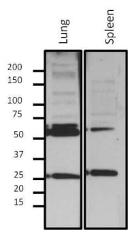
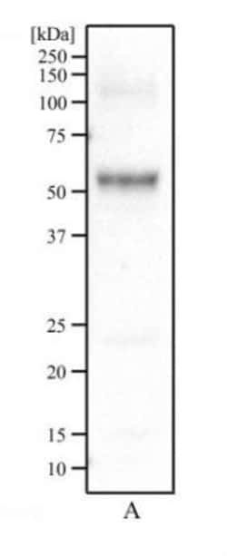
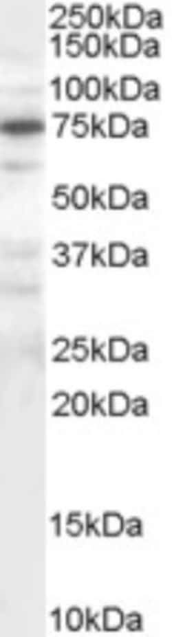
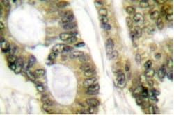
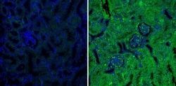
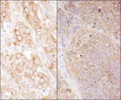
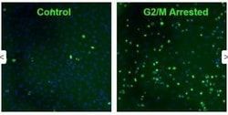
In Western blot this antibody detects ∼17 kDa protein representing phosphorylated H2AX in gamma irradiated HeLa cell lysate. In immunofluorescence procedures, recognizes phosphorylated H2AX in gamma irradiated HeLa cells. ELISA of phosphorylated H2AX can also be performed. Used in IHC to successfully detect H2A.X pSer140 in postnatal mouse lung section.
Content And Storage
Store at -20C. Avoid freeze-thaw cycles.
Isotype
IgG1 κ
Related Products


Description
- Description Histone H2AX (p Ser139) Monoclonal antibody specifically detects Histone H2AX (p Ser139) in Human, Mouse, Bovine samples
- It is validated for Western Blot, Flow Cytometry, ELISA, Immunohistochemistry, Immunocytochemistry, Immunofluorescence, Immunohistochemistry (Paraffin).
Compare Similar Items
Show Difference
Antigen: Histone H2AX (p Ser139)
Classification: Monoclonal
Concentration: 1 mg/mL
Dilution: Western Blot 1 ug/ml, Simple Western 10 ug/ml, Flow Cytometry 1 ug 10^6 cells, ELISA 1:100 - 1:2000, Immunohistochemistry 1:10 - 1:500, Immunocytochemistry/Immunofluorescence 2 - 4 ug/ml, Immunohistochemistry-Paraffin 1:10 - 1:500
Gene Alias: H2A.X, H2A/X, H2AFX
Host Species: Mouse
Molecular Weight of Antigen: 15 kDa
Quantity: 100 μg
Research Discipline: Checkpoint signaling, DNA Double Strand Break Repair, DNA Repair, Epigenetics, Mitotic Regulators, Phospho Specific
Gene ID (Entrez): 3014
Target Species: Human, Mouse, Bovine
Form: Purified
Applications: Western Blot, Flow Cytometry, ELISA, Immunohistochemistry, Immunocytochemistry, Immunofluorescence
Clone: 3F2
Conjugate: Unconjugated
Gene Accession No.: P16104
Gene Symbols: H2AFX
Immunogen: This Histone H2AX [p Ser139] Antibody (3F2) was developed against a synthetic peptide sequence surrounding phosphorylated Ser139.
Purification Method: Protein G purified
Regulatory Status: RUO
Primary or Secondary: Primary
Test Specificity: In Western blot this antibody detects ∼17 kDa protein representing phosphorylated H2AX in gamma irradiated HeLa cell lysate. In immunofluorescence procedures, recognizes phosphorylated H2AX in gamma irradiated HeLa cells. ELISA of phosphorylated H2AX can also be performed. Used in IHC to successfully detect H2A.X pSer140 in postnatal mouse lung section.
Content And Storage: Store at -20C. Avoid freeze-thaw cycles.
Isotype: IgG1 κ
Antigen: HCN4
Classification: Monoclonal
Concentration: __
Dilution: Western Blot 1:5000, Flow Cytometry 1:50, Immunohistochemistry 1:10 - 1:500, Immunohistochemistry-Paraffin 1:10 - 1:100, Immunohistochemistry-Frozen 1:1000, Immunofluorescence 1:10 - 1:500
Gene Alias: hyperpolarization activated cyclic nucleotide-gated cation channel 4, hyperpolarization activated cyclic nucleotide-gated potassium channel 4, potassium/sodium hyperpolarization-activated cyclic nucleotide-gated channel 4, SSS2
Host Species: Rat
Molecular Weight of Antigen: __
Quantity: 100 μL
Research Discipline: __
Gene ID (Entrez): 10021
Target Species: Human, Mouse, Rat
Form: Ascites
Applications: Western Blot, Flow Cytometry, Immunohistochemistry, Immunohistochemistry (Paraffin), Immunohistochemistry (Frozen)
Clone: SHG 1E5
Conjugate: Unconjugated
Gene Accession No.: Q9Y3Q4
Gene Symbols: HCN4
Immunogen: Synthetic peptide sequence corresponding to residues S H G S L L L P P A S S P P P P Q V P Q R R G T P P L T P G R L T Q D L K L of HCN4.
Purification Method: Unpurified
Regulatory Status: RUO
Primary or Secondary: Primary
Test Specificity: HCN4 (SHG 1E5)
Content And Storage: Store at -20C. Avoid freeze-thaw cycles.
Isotype: IgG1
Antigen: IRS1 (p Tyr1179)
Classification: Polyclonal
Concentration: __
Dilution: Western Blot 1:100 - 1:2000
Gene Alias: HIRS-1, insulin receptor substrate 1, IRS-1
Host Species: Rabbit
Molecular Weight of Antigen: __
Quantity: 100 μg
Research Discipline: Cancer, mTOR Pathway, Phospho Specific
Gene ID (Entrez): 3667
Target Species: Mouse
Form: __
Applications: Western Blot
Clone: __
Conjugate: Unconjugated
Gene Accession No.: __
Gene Symbols: IRS1
Immunogen: Synthetic phosphopeptide corresponding to residues L(1173) E N G L N (pY) I D L D L V K(1186) of human IRS-1.
Purification Method: Affinity Purified
Regulatory Status: RUO
Primary or Secondary: Primary
Test Specificity: Detects phosphorylated IRS-1 (Tyr1173) in mouse cells.
Content And Storage: Store at -20C. Avoid freeze-thaw cycles.
Isotype: IgG