MUC1 Antibody (SM3), Alexa Fluor™ 488, Novus Biologicals™
Manufacturer: Novus Biologicals
Select a Size
| Pack Size | SKU | Availability | Price |
|---|---|---|---|
| Each of 1 | NB12022711X-Each-of-1 | In Stock | ₹ 55,358.00 |
NB12022711X - Each of 1
In Stock
Quantity
1
Base Price: ₹ 55,358.00
GST (18%): ₹ 9,964.44
Total Price: ₹ 65,322.44
Antigen
MUC-1
Classification
Monoclonal
Conjugate
Alexa Fluor 488
Formulation
50mM Sodium Borate with 0.05% Sodium Azide
Gene Symbols
MUC1
Immunogen
Hydrogen fluoride deglycosylated milk mucin.
Quantity
0.1 mL
Research Discipline
Cancer, Cellular Markers, Extracellular Matrix, Inflammation, Signal Transduction
Gene ID (Entrez)
4582
Target Species
Human, Mouse
Form
Purified
Applications
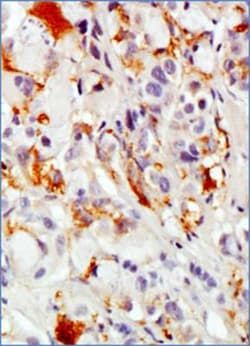
Flow Cytometry, ELISA, Immunohistochemistry, Immunocytochemistry, Immunofluorescence, Immunohistochemistry (Paraffin), Immunohistochemistry (Frozen)
Clone
SM3
Dilution
Flow Cytometry, ELISA, Immunohistochemistry, Immunocytochemistry/Immunofluorescence, Immunohistochemistry-Paraffin, Immunohistochemistry-Frozen
Gene Alias
Breast carcinoma-associated antigen DF3, Carcinoma-associated mucin, CD227, CD227 antigen, DF3 antigen, EMA, episialin, H23 antigen, H23AG, KL-6, MAM6, MUC-1, MUC1/ZD, mucin 1, cell surface associated, mucin 1, transmembrane, mucin-1, Peanut-reactive urinary mucin, PEMMUC-1/SEC, PEMT, Polymorphic epithelial mucin, PUMMUC-1/X, tumor associated epithelial mucin, Tumor-associated epithelial membrane antigen, Tumor-associated mucin
Host Species
Mouse
Purification Method
Protein G purified
Regulatory Status
RUO
Primary or Secondary
Primary
Test Specificity
Reacts very little with normal tissue. Found to react with breast, colon and ovarian carcinomas and adinocarcinoma. SM3 recognises the under-glycosylated form of MUC1 and is therefore tumour specific.
Content And Storage
Store at 4C in the dark.
Isotype
IgG2a κ
Related Products


Description
- MUC1 Monoclonal specifically detects MUC1 in Human, Mouse samples
- It is validated for Flow Cytometry, ELISA, Immunohistochemistry, Immunocytochemistry/Immunofluorescence, Immunohistochemistry-Paraffin, Immunohistochemistry-Frozen.
Compare Similar Items
Show Difference
Antigen: MUC-1
Classification: Monoclonal
Conjugate: Alexa Fluor 488
Formulation: 50mM Sodium Borate with 0.05% Sodium Azide
Gene Symbols: MUC1
Immunogen: Hydrogen fluoride deglycosylated milk mucin.
Quantity: 0.1 mL
Research Discipline: Cancer, Cellular Markers, Extracellular Matrix, Inflammation, Signal Transduction
Gene ID (Entrez): 4582
Target Species: Human, Mouse
Form: Purified
Applications: Flow Cytometry, ELISA, Immunohistochemistry, Immunocytochemistry, Immunofluorescence, Immunohistochemistry (Paraffin), Immunohistochemistry (Frozen)
Clone: SM3
Dilution: Flow Cytometry, ELISA, Immunohistochemistry, Immunocytochemistry/Immunofluorescence, Immunohistochemistry-Paraffin, Immunohistochemistry-Frozen
Gene Alias: Breast carcinoma-associated antigen DF3, Carcinoma-associated mucin, CD227, CD227 antigen, DF3 antigen, EMA, episialin, H23 antigen, H23AG, KL-6, MAM6, MUC-1, MUC1/ZD, mucin 1, cell surface associated, mucin 1, transmembrane, mucin-1, Peanut-reactive urinary mucin, PEMMUC-1/SEC, PEMT, Polymorphic epithelial mucin, PUMMUC-1/X, tumor associated epithelial mucin, Tumor-associated epithelial membrane antigen, Tumor-associated mucin
Host Species: Mouse
Purification Method: Protein G purified
Regulatory Status: RUO
Primary or Secondary: Primary
Test Specificity: Reacts very little with normal tissue. Found to react with breast, colon and ovarian carcinomas and adinocarcinoma. SM3 recognises the under-glycosylated form of MUC1 and is therefore tumour specific.
Content And Storage: Store at 4C in the dark.
Isotype: IgG2a κ
Antigen: MUC-1
Classification: Monoclonal
Conjugate: Alexa Fluor 647
Formulation: 50mM Sodium Borate with 0.05% Sodium Azide
Gene Symbols: MUC1
Immunogen: Hydrogen fluoride deglycosylated milk mucin.
Quantity: 0.1 mL
Research Discipline: Cancer, Cellular Markers, Extracellular Matrix, Inflammation, Signal Transduction
Gene ID (Entrez): 4582
Target Species: Human, Mouse
Form: Purified
Applications: Flow Cytometry, ELISA, Immunohistochemistry, Immunocytochemistry, Immunofluorescence, Immunohistochemistry (Paraffin), Immunohistochemistry (Frozen)
Clone: SM3
Dilution: Flow Cytometry, ELISA, Immunohistochemistry, Immunocytochemistry/Immunofluorescence, Immunohistochemistry-Paraffin, Immunohistochemistry-Frozen
Gene Alias: Breast carcinoma-associated antigen DF3, Carcinoma-associated mucin, CD227, CD227 antigen, DF3 antigen, EMA, episialin, H23 antigen, H23AG, KL-6, MAM6, MUC-1, MUC1/ZD, mucin 1, cell surface associated, mucin 1, transmembrane, mucin-1, Peanut-reactive urinary mucin, PEMMUC-1/SEC, PEMT, Polymorphic epithelial mucin, PUMMUC-1/X, tumor associated epithelial mucin, Tumor-associated epithelial membrane antigen, Tumor-associated mucin
Host Species: Mouse
Purification Method: Protein G purified
Regulatory Status: RUO
Primary or Secondary: Primary
Test Specificity: Reacts very little with normal tissue. Found to react with breast, colon and ovarian carcinomas and adinocarcinoma. SM3 recognises the under-glycosylated form of MUC1 and is therefore tumour specific.
Content And Storage: Store at 4C in the dark.
Isotype: IgG2a κ
Antigen: Integrin alpha 4/CD49d
Classification: Monoclonal
Conjugate: Unconjugated
Formulation: __
Gene Symbols: ITGA4
Immunogen: JM leukaemia line (Human).
Quantity: 0.1 mg
Research Discipline: Cancer, Cellular Markers, Cytoskeleton Markers
Gene ID (Entrez): 3676
Target Species: Human
Form: Purified
Applications: Flow Cytometry, Immunohistochemistry, Immunoprecipitation, Immunohistochemistry (Frozen)
Clone: HP2/1
Dilution: Flow Cytometry 1ug/5 x 10^5 cells, Immunohistochemistry 1:10-1:500, Immunoprecipitation 1:10-1:500, Immunohistochemistry-Frozen 1:10-1:500
Gene Alias: 269C wild type, CD49 antigen-like family member D, CD49d, CD49d antigen, CD49Dantigen CD49D, alpha-4 subunit of VLA-4 receptor, IA4, integrin alpha 4, integrin alpha-4, integrin alpha-4 subunit, Integrin alpha-IV, integrin, alpha 4 (antigen CD49D, alpha 4 subunit of VLA-4 receptor), MGC90518, very late activation protein 4 receptor, alpha 4 subunit, VLA-4 subunit alpha
Host Species: Mouse
Purification Method: Protein A or G purified
Regulatory Status: RUO
Primary or Secondary: Primary
Test Specificity: Mouse anti Human CD49d monoclonal antibody, clone HP2/1 recognizes human CD49d also known as integrin alpha-4 or VLA-4 subunit alpha. CD49d is a ∼150kDa single pass type 1 transmembrane glycoprotein with seven FG-GAP repeats, characteristic of alpha integrins, in its extracellular domain. CD49d can be proteolytically cleaved to yield framents of 80 and 70kDa (Hemler et al. 1987). CD49d associates with either CD29 to form VLA-4 or with Integrin beta-7 to form The Peyer patches-specific homing receptor LPAM-1, involved in the lymphocyte migration and homing to gut-associated lymphoid tissue (Sackstein 2006) through its interaction with MadCam-1, preferentially expressed on Peyer's patch high endothelial venules and postcapillary venules in lamina propria (Briskin et al. 1997). Mouse anti human CD49d, clone HP2/1 binds to both intact and the 80kDa fragment of integrin alpha-4. CD49d is expressed on monocytes, T cells, B cells, thymocytes and Langerhans cells (de Graaf et al. 1995). Mouse anti Human CD49d, clone HP2/1 can be used in basic studies of VLA-4 mediated adhesion and its interaction with the VCAM-1 structure and has been demonstrated to inhibit cell binding to soluble VCAM-1 (Weller et al. 1991).
Content And Storage: Store at 4C short term. Aliquot and store at -20C long term. Avoid freeze-thaw cycles.
Isotype: IgG1