CD63 Antibody (NKI/C3 + LAMP3/968), Alexa Fluor™ 405, Novus Biologicals™
Manufacturer: Novus Biologicals
Select a Size
| Pack Size | SKU | Availability | Price |
|---|---|---|---|
| Each of 1 | NB247937405-Each-of-1 | In Stock | ₹ 58,562.00 |
NB247937405 - Each of 1
In Stock
Quantity
1
Base Price: ₹ 58,562.00
GST (18%): ₹ 10,541.16
Total Price: ₹ 69,103.16
Antigen
CD63
Classification
Monoclonal
Conjugate
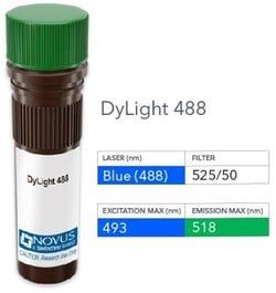
Alexa Fluor 405
Formulation
50mM Sodium Borate with 0.05% Sodium Azide
Gene Symbols
CD63
Immunogen
Smooth plasma membrane fraction of MeWo cells (NKI/C3); Recombinant human full-length CD63 protein (LAMP3/968)
Quantity
0.1 mL
Primary or Secondary
Primary
Test Specificity
This monoclonal antibody recognizes protein of 26kDa-60kDa, which is identified as CD63. The tetraspanins are integral membrane proteins expressed on cell surface and granular membranes of hematopoietic cells and are components of multi-molecular complexes with specific integrins. The tetraspanin CD63 is a lysosomal membrane glycoprotein that translocates to the plasma membrane after platelet activation. CD63 is expressed on activated platelets, monocytes and macrophages, and is weakly expressed on granulocytes, T cell and B cells. It is located on the basophilic granule membranes and on the plasma membranes of lymphocytes and granulocytes. CD63 is a member of the TM4 superfamily of leukocyte glycoproteins that includes CD9, CD37 and CD53, which contain four transmembrane regions. CD63 may play a role in phagocytic and intracellular lysosome-phagosome fusion events. CD63 deficiency is associated with Hermansky-Pudlak syndrome and is strongly expressed during the early stages of melanoma progression.
Content And Storage
Store at 4C in the dark.
Isotype
IgG1 κ
Applications
Flow Cytometry, Immunohistochemistry, Immunohistochemistry (Paraffin), Immunofluorescence
Clone
NKI/C3 + LAMP3/968
Dilution
Flow Cytometry, Immunohistochemistry, Immunohistochemistry-Paraffin, Immunofluorescence
Gene Alias
CD63 antigen, CD63 antigen (melanoma 1 antigen), CD63 molecule, Granulophysin, LAMP-3, Lysosomal-associated membrane protein 3, ME491, melanoma 1 antigen, Melanoma-associated antigen ME491, melanoma-associated antigen MLA1, MLA1lysosome-associated membrane glycoprotein 3, Ocular melanoma-associated antigen, OMA81H, Tetraspanin-30, tspan-30, TSPAN30granulophysin
Host Species
Mouse
Purification Method
Protein A or G purified
Research Discipline
Autophagy, Cancer, Cardiovascular Biology, Cytokine Research, Immunology, Innate Immunity, Lysosome Markers, Metastasis
Gene ID (Entrez)
967
Target Species
Human
Form
Purified
Description
- CD63 Monoclonal specifically detects CD63 in Human samples
- It is validated for Flow Cytometry, Immunohistochemistry, Immunocytochemistry/Immunofluorescence, Immunohistochemistry-Paraffin, Immunofluorescence.
Compare Similar Items
Show Difference

Antigen: CD63
Classification: Monoclonal
Conjugate: Alexa Fluor 405
Formulation: 50mM Sodium Borate with 0.05% Sodium Azide
Gene Symbols: CD63
Immunogen: Smooth plasma membrane fraction of MeWo cells (NKI/C3); Recombinant human full-length CD63 protein (LAMP3/968)
Quantity: 0.1 mL
Primary or Secondary: Primary
Test Specificity: This monoclonal antibody recognizes protein of 26kDa-60kDa, which is identified as CD63. The tetraspanins are integral membrane proteins expressed on cell surface and granular membranes of hematopoietic cells and are components of multi-molecular complexes with specific integrins. The tetraspanin CD63 is a lysosomal membrane glycoprotein that translocates to the plasma membrane after platelet activation. CD63 is expressed on activated platelets, monocytes and macrophages, and is weakly expressed on granulocytes, T cell and B cells. It is located on the basophilic granule membranes and on the plasma membranes of lymphocytes and granulocytes. CD63 is a member of the TM4 superfamily of leukocyte glycoproteins that includes CD9, CD37 and CD53, which contain four transmembrane regions. CD63 may play a role in phagocytic and intracellular lysosome-phagosome fusion events. CD63 deficiency is associated with Hermansky-Pudlak syndrome and is strongly expressed during the early stages of melanoma progression.
Content And Storage: Store at 4C in the dark.
Isotype: IgG1 κ
Applications: Flow Cytometry, Immunohistochemistry, Immunohistochemistry (Paraffin), Immunofluorescence
Clone: NKI/C3 + LAMP3/968
Dilution: Flow Cytometry, Immunohistochemistry, Immunohistochemistry-Paraffin, Immunofluorescence
Gene Alias: CD63 antigen, CD63 antigen (melanoma 1 antigen), CD63 molecule, Granulophysin, LAMP-3, Lysosomal-associated membrane protein 3, ME491, melanoma 1 antigen, Melanoma-associated antigen ME491, melanoma-associated antigen MLA1, MLA1lysosome-associated membrane glycoprotein 3, Ocular melanoma-associated antigen, OMA81H, Tetraspanin-30, tspan-30, TSPAN30granulophysin
Host Species: Mouse
Purification Method: Protein A or G purified
Research Discipline: Autophagy, Cancer, Cardiovascular Biology, Cytokine Research, Immunology, Innate Immunity, Lysosome Markers, Metastasis
Gene ID (Entrez): 967
Target Species: Human
Form: Purified
Antigen: CD63
Classification: Monoclonal
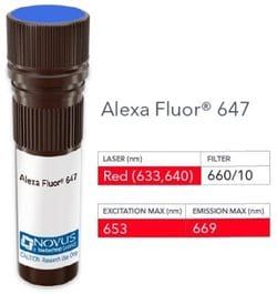
Conjugate: Alexa Fluor 647
Formulation: 50mM Sodium Borate with 0.05% Sodium Azide
Gene Symbols: CD63
Immunogen: Smooth plasma membrane fraction of MeWo cells (NKI/C3); Recombinant human full-length CD63 protein (LAMP3/968)
Quantity: 0.1 mL
Primary or Secondary: Primary
Test Specificity: This monoclonal antibody recognizes protein of 26kDa-60kDa, which is identified as CD63. The tetraspanins are integral membrane proteins expressed on cell surface and granular membranes of hematopoietic cells and are components of multi-molecular complexes with specific integrins. The tetraspanin CD63 is a lysosomal membrane glycoprotein that translocates to the plasma membrane after platelet activation. CD63 is expressed on activated platelets, monocytes and macrophages, and is weakly expressed on granulocytes, T cell and B cells. It is located on the basophilic granule membranes and on the plasma membranes of lymphocytes and granulocytes. CD63 is a member of the TM4 superfamily of leukocyte glycoproteins that includes CD9, CD37 and CD53, which contain four transmembrane regions. CD63 may play a role in phagocytic and intracellular lysosome-phagosome fusion events. CD63 deficiency is associated with Hermansky-Pudlak syndrome and is strongly expressed during the early stages of melanoma progression.
Content And Storage: Store at 4C in the dark.
Isotype: IgG1 κ
Applications: Flow Cytometry, Immunohistochemistry, Immunohistochemistry (Paraffin), Immunofluorescence
Clone: NKI/C3 + LAMP3/968
Dilution: Flow Cytometry, Immunohistochemistry, Immunohistochemistry-Paraffin, Immunofluorescence
Gene Alias: CD63 antigen, CD63 antigen (melanoma 1 antigen), CD63 molecule, Granulophysin, LAMP-3, Lysosomal-associated membrane protein 3, ME491, melanoma 1 antigen, Melanoma-associated antigen ME491, melanoma-associated antigen MLA1, MLA1lysosome-associated membrane glycoprotein 3, Ocular melanoma-associated antigen, OMA81H, Tetraspanin-30, tspan-30, TSPAN30granulophysin
Host Species: Mouse
Purification Method: Protein A or G purified
Research Discipline: Autophagy, Cancer, Cardiovascular Biology, Cytokine Research, Immunology, Innate Immunity, Lysosome Markers, Metastasis
Gene ID (Entrez): 967
Target Species: Human
Form: Purified
Antigen: CD63
Classification: Monoclonal
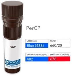
Conjugate: PerCP
Formulation: PBS with 0.05% Sodium Azide
Gene Symbols: CD63
Immunogen: Smooth plasma membrane fraction of MeWo cells (NKI/C3); Recombinant human full-length CD63 protein (LAMP3/968)
Quantity: 0.1 mL
Primary or Secondary: Primary
Test Specificity: This monoclonal antibody recognizes protein of 26kDa-60kDa, which is identified as CD63. The tetraspanins are integral membrane proteins expressed on cell surface and granular membranes of hematopoietic cells and are components of multi-molecular complexes with specific integrins. The tetraspanin CD63 is a lysosomal membrane glycoprotein that translocates to the plasma membrane after platelet activation. CD63 is expressed on activated platelets, monocytes and macrophages, and is weakly expressed on granulocytes, T cell and B cells. It is located on the basophilic granule membranes and on the plasma membranes of lymphocytes and granulocytes. CD63 is a member of the TM4 superfamily of leukocyte glycoproteins that includes CD9, CD37 and CD53, which contain four transmembrane regions. CD63 may play a role in phagocytic and intracellular lysosome-phagosome fusion events. CD63 deficiency is associated with Hermansky-Pudlak syndrome and is strongly expressed during the early stages of melanoma progression.
Content And Storage: Store at 4C in the dark.
Isotype: IgG1 κ
Applications: Flow Cytometry
Clone: NKI/C3 + LAMP3/968
Dilution: Flow Cytometry
Gene Alias: CD63 antigen, CD63 antigen (melanoma 1 antigen), CD63 molecule, Granulophysin, LAMP-3, Lysosomal-associated membrane protein 3, ME491, melanoma 1 antigen, Melanoma-associated antigen ME491, melanoma-associated antigen MLA1, MLA1lysosome-associated membrane glycoprotein 3, Ocular melanoma-associated antigen, OMA81H, Tetraspanin-30, tspan-30, TSPAN30granulophysin
Host Species: Mouse
Purification Method: Protein A or G purified
Research Discipline: Autophagy, Cancer, Cardiovascular Biology, Cytokine Research, Immunology, Innate Immunity, Lysosome Markers, Metastasis
Gene ID (Entrez): 967
Target Species: Human
Form: Purified