anti-IL-17/IL-17A, HRP, Polyclonal, Novus Biologicals™
Manufacturer: Novus Biologicals
Select a Size
| Pack Size | SKU | Availability | Price |
|---|---|---|---|
| Each of 1 | NBP142774-Each-of-1 | In Stock | ₹ 59,719.00 |
NBP142774 - Each of 1
In Stock
Quantity
1
Base Price: ₹ 59,719.00
GST (18%): ₹ 10,749.42
Total Price: ₹ 70,468.42
Antigen
IL-17/IL-17A
Classification
Polyclonal
Conjugate
HRP
Formulation
Lyophilized from 0.02 M Potassium Phosphate, 0.15 M Sodium Chloride, pH 7.2, 10 mg/mL Bovine Serum Albumin (BSA) - Immunoglobulin and Protease free with 0.01% Gentamicin Sulfate
Gene Symbols
IL17A
Immunogen
This purified IL-17/IL-17A Antibody was prepared from whole rabbit serum produced by repeated immunizations with full length recombinant rat IL-17/IL-17A protein. (Uniprot: Q61453)
Quantity
0.1 mg
Research Discipline
Apoptosis, Cytokine Research, Immunology, Innate Immunity
Gene ID (Entrez)
3605
Reconstitution
Reconstitute with 100 ul deionized water (or equivalent)
Content And Storage
Store lyophilized antibody at 4°C. Aliquot reconstituted liquid and store at -20°C. Avoid freeze-thaw cycles.
Applications
Western Blot, ELISA, Immunohistochemistry
Concentration
LYOPH
Dilution
Western Blot 1:1000-1:5000, ELISA 1:10000-1:50000, Immunohistochemistry 1:500-1:2500
Gene Alias
CTLA8cytotoxic T-lymphocyte-associated serine esterase 8, Cytotoxic T-lymphocyte-associated antigen 8, IL-17Acytotoxic T-lymphocyte-associated protein 8, IL-17CTLA-8, IL17interleukin-17A, interleukin 17 (cytotoxic T-lymphocyte-associated serine esterase 8), interleukin 17A
Host Species
Rabbit
Purification Method
Affinity Purified
Regulatory Status
RUO
Primary or Secondary
Primary
Test Specificity
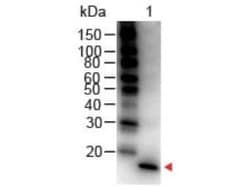
In ELISA and other immunoreactive assays, this antibody will recognize both native and recombinant rat IL-17/IL-17A in cell supernatants and certain body fluids. A control of similarly diluted normal rabbit IgG is recommended.
Target Species
Human, Mouse, Rat
Isotype
IgG
Related Products


Description
- IL-17/IL-17A Polyclonal specifically detects IL-17/IL-17A in Human, Mouse, Rat samples
- It is validated for Western Blot, ELISA, Immunohistochemistry.
Compare Similar Items
Show Difference

Antigen: IL-17/IL-17A
Classification: Polyclonal
Conjugate: HRP
Formulation: Lyophilized from 0.02 M Potassium Phosphate, 0.15 M Sodium Chloride, pH 7.2, 10 mg/mL Bovine Serum Albumin (BSA) - Immunoglobulin and Protease free with 0.01% Gentamicin Sulfate
Gene Symbols: IL17A
Immunogen: This purified IL-17/IL-17A Antibody was prepared from whole rabbit serum produced by repeated immunizations with full length recombinant rat IL-17/IL-17A protein. (Uniprot: Q61453)
Quantity: 0.1 mg
Research Discipline: Apoptosis, Cytokine Research, Immunology, Innate Immunity
Gene ID (Entrez): 3605
Reconstitution: Reconstitute with 100 ul deionized water (or equivalent)
Content And Storage: Store lyophilized antibody at 4°C. Aliquot reconstituted liquid and store at -20°C. Avoid freeze-thaw cycles.
Applications: Western Blot, ELISA, Immunohistochemistry
Concentration: LYOPH
Dilution: Western Blot 1:1000-1:5000, ELISA 1:10000-1:50000, Immunohistochemistry 1:500-1:2500
Gene Alias: CTLA8cytotoxic T-lymphocyte-associated serine esterase 8, Cytotoxic T-lymphocyte-associated antigen 8, IL-17Acytotoxic T-lymphocyte-associated protein 8, IL-17CTLA-8, IL17interleukin-17A, interleukin 17 (cytotoxic T-lymphocyte-associated serine esterase 8), interleukin 17A
Host Species: Rabbit
Purification Method: Affinity Purified
Regulatory Status: RUO
Primary or Secondary: Primary
Test Specificity: In ELISA and other immunoreactive assays, this antibody will recognize both native and recombinant rat IL-17/IL-17A in cell supernatants and certain body fluids. A control of similarly diluted normal rabbit IgG is recommended.
Target Species: Human, Mouse, Rat
Isotype: IgG

Antigen: His Tag
Classification: Monoclonal
Conjugate: HRP
Formulation: __
Gene Symbols: __
Immunogen: His Tag Antibody (33D10.D2.G8) was produced in mice by repeated immunizations with 6X His epitope tag peptide H-H-H-H-H-H conjugated to KLH using maleimide.
Quantity: 0.1 mg
Research Discipline: Cellular Markers, Epitope Tags
Gene ID (Entrez): __
Reconstitution: Reconstitute with 100 ul deionized water (or equivalent)
Content And Storage: Store lyophilized antibody at 4°C. Aliquot reconstituted liquid and store at -20°C. Avoid freeze-thaw cycles.
Applications: Western Blot, ELISA, Immunohistochemistry, Immunohistochemistry (Paraffin)
Concentration: LYOPH
Dilution: Western Blot 1:1000-1:5000, ELISA 1:10000, Immunohistochemistry 1:500-1:2500, Immunohistochemistry-Paraffin 1:10-1:500
Gene Alias: 6 His epitope tag, 6X His, 6X-His, H, HHHHHH epitope tag, HHHHHH tag, HIS, His tag, Poly-histidine
Host Species: Mouse
Purification Method: Affinity Purified
Regulatory Status: RUO
Primary or Secondary: Primary
Test Specificity: This protein-A purified antibody is directed against the 6X His motif and is useful in determining its presence in various assays. This monoclonal anti-6X His tag antibody detects over-expressed proteins containing the 6X His epitope tag. To date, this antibody has reacted with all His tagged proteins so far tested. In western blotting of bacterial extracts, the antibody does not cross-react with endogenous proteins. The antibody recognizes the His-tag (His-His-His-His-His-His) fused to either the amino- or carboxy-termini of targeted proteins in transfected or transformed cells.
Target Species: __
Isotype: IgG1 κ

Antigen: L-Selectin/CD62L
Classification: Monoclonal
Conjugate: FITC
Formulation: __
Gene Symbols: SELL
Immunogen: PMA-activated human peripheral blood leukocytes
Quantity: 100 Tests
Research Discipline: Adaptive Immunity, B Cell Development and Differentiation Markers, Cardiovascular Biology, Glycobiology, Immunology, Lipid and Metabolism, Myeloid derived Suppressor Cell, Signal Transduction, Stem Cells
Gene ID (Entrez): 6402
Reconstitution: __
Content And Storage: Store at 4C in the dark.
Applications: Western Blot, Flow Cytometry, Immunohistochemistry, Immunoprecipitation, Immunohistochemistry (Frozen), Functional Assay
Concentration: __
Dilution: Western Blot 1:100-1:2000, Flow Cytometry, Immunohistochemistry 1:10-1:500, Immunoprecipitation 1:10-1:500, Immunohistochemistry-Frozen 1:10-1:500, Functional
Gene Alias: CD62L, CD62L antigen, gp90-MEL, hLHRc, LAM-1, LAM1LECAM1, Leu-8, LEU8, Leukocyte adhesion molecule 1, Leukocyte surface antigen Leu-8, Leukocyte-endothelial cell adhesion molecule 1, LNHRTQ1, LSEL, L-selectin, Lyam-1, LYAM1CD62 antigen-like family member L, Lymph node homing receptor, lymphocyte adhesion molecule 1, pln homing receptor, PLNHR, selectin L
Host Species: Mouse
Purification Method: Size Exclusion Chromatography
Regulatory Status: RUO
Primary or Secondary: Primary
Test Specificity: The mouse monoclonal antibody DREG56 recognizes CD62L L-selectin, a 65-76 kDa cell surface protein, expressed by neutrophils, monocytes, and subsets of T, B, and NK cells, that interacts with specific carbohydrates exposed on activated endothelial cells. HLDA V; WS Code S056
Target Species: Human
Isotype: IgG1