Astrocytomas Antibody (J1-31), Novus Biologicals™
Manufacturer: Fischer Scientific
Select a Size
| Pack Size | SKU | Availability | Price |
|---|---|---|---|
| Each of 1 | NBP229820-Each-of-1 | In Stock | ₹ 61,187.50 |
NBP229820 - Each of 1
In Stock
Quantity
1
Base Price: ₹ 61,187.50
GST (18%): ₹ 11,013.75
Total Price: ₹ 72,201.25
Antigen
Astrocytomas
Classification
Monoclonal
Conjugate
Unconjugated
Formulation
Ascites with No Preservative
Host Species
Mouse
Purification Method
Unpurified
Regulatory Status
RUO
Test Specificity
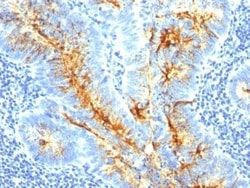
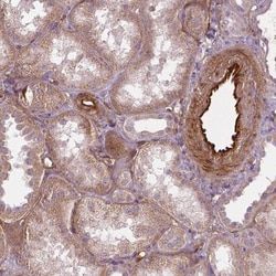
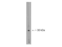
The antibody recognizes an intracellular protein antigen (MW 30 kDa) expressed by human and rat astrocytes and other specialized glia (Muller cells of the retina, Bergmann fibers of the cerebellar cortex, tanycytes of the hypothalamus and ciliated ependymal cells) in the central nervous system (CNS).The antibody has recently been found to be a specific marker for low grade astrocytoma in human brain tissue. The antibody is able to distinguish between low grade astrocytoma and normal reactive gliosis (patent application filed). Monoclonal antibody J1-31 was raised against crude homogenate of brain tissue from a multiple sclerosis (MS) patient (autopsy sample; Malhotra et al.: Microbios Letters 26:151-157, 1984). In human brain, MAb J1-31 recognizes an intracellular protein antigen (J1-31 antigen), which bands at approximately 30,000 daltons under reducing conditions for sodium dodecyl sulfate gel electrophoresis (Singh et al.: Bioscience Reports 6:73-79, 1986). By immunofluorescence microscopy, MAb J1-31 stains those cells that are also stained by antiserum to glial fibrillary acidic protein (GFAP), namely astrocytes, retinal Muller cells, and tanycytes in the ependyma (Predy et al.: Bioscience Reports 7:491-502, 1987). In addition, MAb J1-31 stains ciliated ependymal cells that do not express GFAP (Malhotra, SK (1989) J Neurosci Res. 22(1):36-49).
Content And Storage
Aliquot and store at -20C or -80C. Avoid freeze-thaw cycles.
Isotype
IgM
Applications
Western Blot, Immunocytochemistry, Immunofluorescence, Immunohistochemistry (Paraffin)
Clone
J1-31
Dilution
Western Blot 1:100-1:2000, Immunocytochemistry/Immunofluorescence 1:10-1:500, Immunohistochemistry-Paraffin 1:10-1:500
Gene Alias
Astrocytoma Marker, Astrocytomas Marker, Low Grade Astrocytoma Marker
Immunogen
Human cerebral white matter plaque materials from a multiple sclerosis patient.
Quantity
0.1 mL
Primary or Secondary
Primary
Target Species
Human, Rat
Form
Ascites
Related Products


Description
- Astrocytomas Monoclonal specifically detects Astrocytomas in Human, Rat samples
- It is validated for Western Blot, Immunohistochemistry, Immunocytochemistry/Immunofluorescence, Immunohistochemistry-Paraffin.
Compare Similar Items
Show Difference

Antigen: Astrocytomas
Classification: Monoclonal
Conjugate: Unconjugated
Formulation: Ascites with No Preservative
Host Species: Mouse
Purification Method: Unpurified
Regulatory Status: RUO
Test Specificity: The antibody recognizes an intracellular protein antigen (MW 30 kDa) expressed by human and rat astrocytes and other specialized glia (Muller cells of the retina, Bergmann fibers of the cerebellar cortex, tanycytes of the hypothalamus and ciliated ependymal cells) in the central nervous system (CNS).The antibody has recently been found to be a specific marker for low grade astrocytoma in human brain tissue. The antibody is able to distinguish between low grade astrocytoma and normal reactive gliosis (patent application filed). Monoclonal antibody J1-31 was raised against crude homogenate of brain tissue from a multiple sclerosis (MS) patient (autopsy sample; Malhotra et al.: Microbios Letters 26:151-157, 1984). In human brain, MAb J1-31 recognizes an intracellular protein antigen (J1-31 antigen), which bands at approximately 30,000 daltons under reducing conditions for sodium dodecyl sulfate gel electrophoresis (Singh et al.: Bioscience Reports 6:73-79, 1986). By immunofluorescence microscopy, MAb J1-31 stains those cells that are also stained by antiserum to glial fibrillary acidic protein (GFAP), namely astrocytes, retinal Muller cells, and tanycytes in the ependyma (Predy et al.: Bioscience Reports 7:491-502, 1987). In addition, MAb J1-31 stains ciliated ependymal cells that do not express GFAP (Malhotra, SK (1989) J Neurosci Res. 22(1):36-49).
Content And Storage: Aliquot and store at -20C or -80C. Avoid freeze-thaw cycles.
Isotype: IgM
Applications: Western Blot, Immunocytochemistry, Immunofluorescence, Immunohistochemistry (Paraffin)
Clone: J1-31
Dilution: Western Blot 1:100-1:2000, Immunocytochemistry/Immunofluorescence 1:10-1:500, Immunohistochemistry-Paraffin 1:10-1:500
Gene Alias: Astrocytoma Marker, Astrocytomas Marker, Low Grade Astrocytoma Marker
Immunogen: Human cerebral white matter plaque materials from a multiple sclerosis patient.
Quantity: 0.1 mL
Primary or Secondary: Primary
Target Species: Human, Rat
Form: Ascites
Antigen: Opsin 1 (Medium Wave)
Classification: Polyclonal
Conjugate: Unconjugated
Formulation: PBS with 0.02% Sodium Azide
Host Species: Chicken
Purification Method: Affinity Purified
Regulatory Status: RUO
Test Specificity: Opsin, red and green. The antibody reacts with a protein of ∼40 kDa.
Content And Storage: Aliquot and store at -20C or -80C. Avoid freeze-thaw cycles.
Isotype: __
Applications: Western Blot, ELISA
Clone: __
Dilution: Western Blot 1:500:1:2000, ELISA 1:100-1:2000
Gene Alias: GCP, GOP, Green cone photoreceptor pigment, Green-sensitive opsin, medium-wave-sensitive opsin 1, OPN1MW, opsin 1 (cone pigments), medium-wave-sensitive 2
Immunogen: Synthetic peptides from mouse green opsin and zebra finch red opsin.
Quantity: 0.1 mg
Primary or Secondary: Primary
Target Species: Mouse, Rat
Form: __
Antigen: LBX1
Classification: Polyclonal
Conjugate: Unconjugated
Formulation: PBS with 0.1% Sodium Azide
Host Species: Rabbit
Purification Method: Affinity Purified
Regulatory Status: RUO
Test Specificity: Lbx1 (Ladybird). By Western Blot the antibody recognizes a band at ∼30 kDa on mouse spinal cord lysate.
Content And Storage: Aliquot and store at -20C or -80C. Avoid freeze-thaw cycles.
Isotype: IgG
Applications: Western Blot
Clone: __
Dilution: Western Blot 1:500-1:1000
Gene Alias: homeobox, HPX6, HPX-6, ladybird homeobox 1, ladybird homeobox homolog 1, ladybird homeobox homolog 1 (Drosophila), Ladybird homeobox protein homolog 1, LBX1Hlady bird-like homeobox, transcription factor LBX1, transcription factor similar to D. melanogaster homeodomain protein lady birdlate
Immunogen: Synthetic peptide.
Quantity: 0.1 mg
Primary or Secondary: Primary
Target Species: Mouse
Form: __