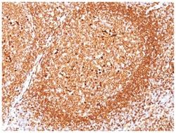
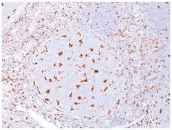
CD68/SR-D1 Mouse, Clone: C68/684, Novus Biologicals™
Manufacturer: Fischer Scientific
Select a Size
| Pack Size | SKU | Availability | Price |
|---|---|---|---|
| Each of 1 | NBP232832A-Each-of-1 | In Stock | ₹ 47,704.00 |
NBP232832A - Each of 1
In Stock
Quantity
1
Base Price: ₹ 47,704.00
GST (18%): ₹ 8,586.72
Total Price: ₹ 56,290.72
Antigen
CD68/SR-D1
Classification
Monoclonal
Conjugate
Unconjugated
Formulation
PBS with 0.05% BSA. with 0.05% Sodium Azide
Gene Alias
CD68 antigenmacrophage antigen CD68, CD68 molecule, DKFZp686M18236, GP110, macrosialin, SCARD1, scavenger receptor class D, member 1
Host Species
Mouse
Molecular Weight of Antigen
110 kDa
Quantity
0.1 mg
Research Discipline
Cell Biology, Cytokine Research, Immunology
Gene ID (Entrez)
968
Target Species
Human, Mouse, Feline, Primate, Rat (Negative)
Form
Purified
Applications
Western Blot, Flow Cytometry, Immunocytochemistry, Immunofluorescence, Immunoprecipitation, Immunohistochemistry (Paraffin)
Clone
C68/684
Dilution
Western Blot 0.5-1ug/ml, Flow Cytometry 0.5-1ug/million cells, Immunocytochemistry/Immunofluorescence 0.5-1ug/ml, Immunoprecipitation 0.5-1ug/500ug protein lysate, Immunohistochemistry-Paraffin 0.5-1.0ug/ml, Immunohistochemistry-Frozen 0.5-1.0ug/ml
Gene Accession No.
P34810
Gene Symbols
CD68
Immunogen
Subcellular fraction of human alveolar macrophages
Purification Method
Protein A purified
Regulatory Status
RUO
Primary or Secondary
Primary
Test Specificity
This antibody recognizes a glycoprotein of 110kDa, which is identified as CD68. It is important for identifying macrophages in tissue sections. It stains macrophages in a wide variety of human tissues, including Kupffer cells and macrophages in the red pulp of the spleen, in lamina propria of the gut, in lung alveoli, and in bone marrow. It reacts with myeloid precursors and peripheral blood granulocytes. It also reacts with plasmacytoid T cells, which are supposed to be of monocyte/macrophage origin. It shows strong granular cytoplasmic staining of chronic and acute myeloid leukemia and also reacts with rare cases of true histiocytic neoplasia. Lymphomas are negative or show few granules.
Content And Storage
Store at 4C.
Isotype
IgG1 κ
Description
- CD68/SR-D1 Monoclonal specifically detects CD68/SR-D1 in Human, Mouse, Rat, Monkey samples
- It is validated for Western Blot, Flow Cytometry, Immunohistochemistry, Immunocytochemistry/Immunofluorescence, Immunohistochemistry-Paraffin, Flow (Intracellular).
Compare Similar Items
Show Difference
Antigen: CD68/SR-D1
Classification: Monoclonal
Conjugate: Unconjugated
Formulation: PBS with 0.05% BSA. with 0.05% Sodium Azide
Gene Alias: CD68 antigenmacrophage antigen CD68, CD68 molecule, DKFZp686M18236, GP110, macrosialin, SCARD1, scavenger receptor class D, member 1
Host Species: Mouse
Molecular Weight of Antigen: 110 kDa
Quantity: 0.1 mg
Research Discipline: Cell Biology, Cytokine Research, Immunology
Gene ID (Entrez): 968
Target Species: Human, Mouse, Feline, Primate, Rat (Negative)
Form: Purified
Applications: Western Blot, Flow Cytometry, Immunocytochemistry, Immunofluorescence, Immunoprecipitation, Immunohistochemistry (Paraffin)
Clone: C68/684
Dilution: Western Blot 0.5-1ug/ml, Flow Cytometry 0.5-1ug/million cells, Immunocytochemistry/Immunofluorescence 0.5-1ug/ml, Immunoprecipitation 0.5-1ug/500ug protein lysate, Immunohistochemistry-Paraffin 0.5-1.0ug/ml, Immunohistochemistry-Frozen 0.5-1.0ug/ml
Gene Accession No.: P34810
Gene Symbols: CD68
Immunogen: Subcellular fraction of human alveolar macrophages
Purification Method: Protein A purified
Regulatory Status: RUO
Primary or Secondary: Primary
Test Specificity: This antibody recognizes a glycoprotein of 110kDa, which is identified as CD68. It is important for identifying macrophages in tissue sections. It stains macrophages in a wide variety of human tissues, including Kupffer cells and macrophages in the red pulp of the spleen, in lamina propria of the gut, in lung alveoli, and in bone marrow. It reacts with myeloid precursors and peripheral blood granulocytes. It also reacts with plasmacytoid T cells, which are supposed to be of monocyte/macrophage origin. It shows strong granular cytoplasmic staining of chronic and acute myeloid leukemia and also reacts with rare cases of true histiocytic neoplasia. Lymphomas are negative or show few granules.
Content And Storage: Store at 4C.
Isotype: IgG1 κ
Antigen: CD68/SR-D1
Classification: Monoclonal
Conjugate: Unconjugated
Formulation: PBS with 0.05% BSA. with 0.05% Sodium Azide
Gene Alias: CD68 antigenmacrophage antigen CD68, CD68 molecule, DKFZp686M18236, GP110, macrosialin, SCARD1, scavenger receptor class D, member 1
Host Species: Mouse
Molecular Weight of Antigen: 110 kDa
Quantity: 0.2 mg
Research Discipline: Cell Biology, Cytokine Research, Immunology
Gene ID (Entrez): 968
Target Species: Human, Mouse, Feline, Primate, Rat (Negative)
Form: Purified
Applications: Western Blot, Flow Cytometry, Immunocytochemistry, Immunofluorescence, Immunoprecipitation, Immunohistochemistry (Paraffin)
Clone: C68/684
Dilution: Western Blot 0.5-1ug/ml, Flow Cytometry 0.5-1ug/million cells, Immunocytochemistry/Immunofluorescence 0.5-1ug/ml, Immunoprecipitation 0.5-1ug/500ug protein lysate, Immunohistochemistry-Paraffin 0.5-1.0ug/ml, Immunohistochemistry-Frozen 0.5-1.0ug/ml
Gene Accession No.: P34810
Gene Symbols: CD68
Immunogen: Subcellular fraction of human alveolar macrophages
Purification Method: Protein A purified
Regulatory Status: RUO
Primary or Secondary: Primary
Test Specificity: This antibody recognizes a glycoprotein of 110kDa, which is identified as CD68. It is important for identifying macrophages in tissue sections. It stains macrophages in a wide variety of human tissues, including Kupffer cells and macrophages in the red pulp of the spleen, in lamina propria of the gut, in lung alveoli, and in bone marrow. It reacts with myeloid precursors and peripheral blood granulocytes. It also reacts with plasmacytoid T cells, which are supposed to be of monocyte/macrophage origin. It shows strong granular cytoplasmic staining of chronic and acute myeloid leukemia and also reacts with rare cases of true histiocytic neoplasia. Lymphomas are negative or show few granules.
Content And Storage: Store at 4C.
Isotype: IgG1 κ
Antigen: CD68/SR-D1
Classification: Monoclonal
Conjugate: Unconjugated
Formulation: PBS with 0.05% BSA. with 0.05% Sodium Azide
Gene Alias: CD68 antigenmacrophage antigen CD68, CD68 molecule, DKFZp686M18236, GP110, macrosialin, SCARD1, scavenger receptor class D, member 1
Host Species: Mouse
Molecular Weight of Antigen: 110 kDa
Quantity: 0.02 mg
Research Discipline: Cell Biology, Cytokine Research, Immunology
Gene ID (Entrez): 968
Target Species: Human, Mouse, Feline, Primate, Rat (Negative)
Form: Purified
Applications: Western Blot, Flow Cytometry, Immunocytochemistry, Immunofluorescence, Immunoprecipitation, Immunohistochemistry (Paraffin)
Clone: C68/684
Dilution: Western Blot 0.5-1ug/ml, Flow Cytometry 0.5-1ug/million cells, Immunocytochemistry/Immunofluorescence 0.5-1ug/ml, Immunoprecipitation 0.5-1ug/500ug protein lysate, Immunohistochemistry-Paraffin 0.5-1.0ug/ml, Immunohistochemistry-Frozen 0.5-1.0ug/ml
Gene Accession No.: P34810
Gene Symbols: CD68
Immunogen: Subcellular fraction of human alveolar macrophages
Purification Method: Protein A purified
Regulatory Status: RUO
Primary or Secondary: Primary
Test Specificity: This antibody recognizes a glycoprotein of 110kDa, which is identified as CD68. It is important for identifying macrophages in tissue sections. It stains macrophages in a wide variety of human tissues, including Kupffer cells and macrophages in the red pulp of the spleen, in lamina propria of the gut, in lung alveoli, and in bone marrow. It reacts with myeloid precursors and peripheral blood granulocytes. It also reacts with plasmacytoid T cells, which are supposed to be of monocyte/macrophage origin. It shows strong granular cytoplasmic staining of chronic and acute myeloid leukemia and also reacts with rare cases of true histiocytic neoplasia. Lymphomas are negative or show few granules.
Content And Storage: Store at 4C.
Isotype: IgG1 κ