Fibronectin/Anastellin Mouse, Clone: SPM246, Novus Biologicals™
Manufacturer: Fischer Scientific
Select a Size
| Pack Size | SKU | Availability | Price |
|---|---|---|---|
| Each of 1 | NBP232850A-Each-of-1 | In Stock | ₹ 50,952.50 |
NBP232850A - Each of 1
In Stock
Quantity
1
Base Price: ₹ 50,952.50
GST (18%): ₹ 9,171.45
Total Price: ₹ 60,123.95
Antigen
Fibronectin
Classification
Monoclonal
Conjugate
Unconjugated
Formulation
PBS with 0.05% BSA. with 0.05% Sodium Azide
Gene Alias
CIGDKFZp686O13149, Cold-insoluble globulin, DKFZp686F10164, DKFZp686H0342, DKFZp686I1370, fibronectin, fibronectin 1, FNFINC, FNZ, GFND2ED-B, LETSGFND, migration-stimulating factor, MSF
Host Species
Mouse
Purification Method
Protein A purified
Regulatory Status
RUO
Primary or Secondary
Primary
Test Specificity
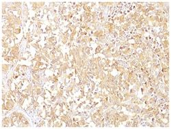
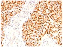
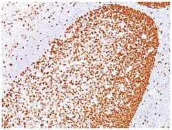
Fibronectin is a soluble dimeric glycoprotein of 440kDa, which is present in cells, extracellular matrix, and blood. This MAb reacts with the cellular as well as plasma form of fibronectin. Reportedly, after iv administration, this MAb localizes to tumor vessels where it binds to the underlying basement. Epitope recognized by this antibody is not accessible in normal tissues to the circulating MAb indicating that it can be used to specifically target tumor vessels in vivo. TV-1 is reportedly useful for delivering vasoactive agents to tumors to induce increased vascular permeability or blood flow prior to treatment with chemotherapeutic drugs or MAbs.
Content And Storage
Store at 4C.
Isotype
IgG1 κ
Applications
Western Blot, Flow Cytometry, Immunocytochemistry, Immunofluorescence, Immunoprecipitation, Immunohistochemistry (Paraffin)
Clone
SPM246
Dilution
Western Blot 0.5-1ug/ml, Flow Cytometry 0.5-1ug/million cells, Immunocytochemistry/Immunofluorescence 0.5-1ug/ml, Immunoprecipitation 0.5-1ug/500ug protein lysate, Immunohistochemistry-Paraffin 0.5-1.0ug/ml, Immunohistochemistry-Frozen 0.5-1.0ug/ml
Gene Accession No.
P02751
Gene Symbols
FN1
Immunogen
T-cell lymphoma biopsy
Quantity
0.1 mg
Research Discipline
Cancer, Cellular Markers, Extracellular Matrix, Mesenchymal Stem Cell Markers, Neuronal Stem Cell Markers, Stem Cell Markers
Gene ID (Entrez)
2335
Target Species
Human, Mouse, Rat, Porcine
Form
Purified
Related Products

Description
- Fibronectin Monoclonal specifically detects Fibronectin in Human, Mouse, Rat, Porcine samples
- It is validated for Flow Cytometry, Immunohistochemistry, Immunocytochemistry/Immunofluorescence, Immunohistochemistry-Paraffin.
Compare Similar Items
Show Difference
Antigen: Fibronectin
Classification: Monoclonal
Conjugate: Unconjugated
Formulation: PBS with 0.05% BSA. with 0.05% Sodium Azide
Gene Alias: CIGDKFZp686O13149, Cold-insoluble globulin, DKFZp686F10164, DKFZp686H0342, DKFZp686I1370, fibronectin, fibronectin 1, FNFINC, FNZ, GFND2ED-B, LETSGFND, migration-stimulating factor, MSF
Host Species: Mouse
Purification Method: Protein A purified
Regulatory Status: RUO
Primary or Secondary: Primary
Test Specificity: Fibronectin is a soluble dimeric glycoprotein of 440kDa, which is present in cells, extracellular matrix, and blood. This MAb reacts with the cellular as well as plasma form of fibronectin. Reportedly, after iv administration, this MAb localizes to tumor vessels where it binds to the underlying basement. Epitope recognized by this antibody is not accessible in normal tissues to the circulating MAb indicating that it can be used to specifically target tumor vessels in vivo. TV-1 is reportedly useful for delivering vasoactive agents to tumors to induce increased vascular permeability or blood flow prior to treatment with chemotherapeutic drugs or MAbs.
Content And Storage: Store at 4C.
Isotype: IgG1 κ
Applications: Western Blot, Flow Cytometry, Immunocytochemistry, Immunofluorescence, Immunoprecipitation, Immunohistochemistry (Paraffin)
Clone: SPM246
Dilution: Western Blot 0.5-1ug/ml, Flow Cytometry 0.5-1ug/million cells, Immunocytochemistry/Immunofluorescence 0.5-1ug/ml, Immunoprecipitation 0.5-1ug/500ug protein lysate, Immunohistochemistry-Paraffin 0.5-1.0ug/ml, Immunohistochemistry-Frozen 0.5-1.0ug/ml
Gene Accession No.: P02751
Gene Symbols: FN1
Immunogen: T-cell lymphoma biopsy
Quantity: 0.1 mg
Research Discipline: Cancer, Cellular Markers, Extracellular Matrix, Mesenchymal Stem Cell Markers, Neuronal Stem Cell Markers, Stem Cell Markers
Gene ID (Entrez): 2335
Target Species: Human, Mouse, Rat, Porcine
Form: Purified
Antigen: Fibronectin
Classification: Monoclonal
Conjugate: Unconjugated
Formulation: PBS with 0.05% BSA. with 0.05% Sodium Azide
Gene Alias: CIGDKFZp686O13149, Cold-insoluble globulin, DKFZp686F10164, DKFZp686H0342, DKFZp686I1370, fibronectin, fibronectin 1, FNFINC, FNZ, GFND2ED-B, LETSGFND, migration-stimulating factor, MSF
Host Species: Mouse
Purification Method: Protein A purified
Regulatory Status: RUO
Primary or Secondary: Primary
Test Specificity: Fibronectin is a soluble dimeric glycoprotein of 440kDa, which is present in cells, extracellular matrix, and blood. This MAb reacts with the cellular as well as plasma form of fibronectin. Reportedly, after iv administration, this MAb localizes to tumor vessels where it binds to the underlying basement. Epitope recognized by this antibody is not accessible in normal tissues to the circulating MAb indicating that it can be used to specifically target tumor vessels in vivo. TV-1 is reportedly useful for delivering vasoactive agents to tumors to induce increased vascular permeability or blood flow prior to treatment with chemotherapeutic drugs or MAbs.
Content And Storage: Store at 4C.
Isotype: IgG1 κ
Applications: Western Blot, Flow Cytometry, Immunocytochemistry, Immunofluorescence, Immunoprecipitation, Immunohistochemistry (Paraffin)
Clone: SPM246
Dilution: Western Blot 0.5-1ug/ml, Flow Cytometry 0.5-1ug/million cells, Immunocytochemistry/Immunofluorescence 0.5-1ug/ml, Immunoprecipitation 0.5-1ug/500ug protein lysate, Immunohistochemistry-Paraffin 0.5-1.0ug/ml, Immunohistochemistry-Frozen 0.5-1.0ug/ml
Gene Accession No.: P02751
Gene Symbols: FN1
Immunogen: T-cell lymphoma biopsy
Quantity: 0.2 mg
Research Discipline: Cancer, Cellular Markers, Extracellular Matrix, Mesenchymal Stem Cell Markers, Neuronal Stem Cell Markers, Stem Cell Markers
Gene ID (Entrez): 2335
Target Species: Human, Mouse, Rat, Porcine
Form: Purified
Antigen: Fibronectin
Classification: Monoclonal
Conjugate: Unconjugated
Formulation: PBS with 0.05% BSA. with 0.05% Sodium Azide
Gene Alias: CIGDKFZp686O13149, Cold-insoluble globulin, DKFZp686F10164, DKFZp686H0342, DKFZp686I1370, fibronectin, fibronectin 1, FNFINC, FNZ, GFND2ED-B, LETSGFND, migration-stimulating factor, MSF
Host Species: Mouse
Purification Method: Protein A purified
Regulatory Status: RUO
Primary or Secondary: Primary
Test Specificity: Fibronectin is a soluble dimeric glycoprotein of 440kDa, which is present in cells, extracellular matrix, and blood. This MAb reacts with the cellular as well as plasma form of fibronectin. Reportedly, after iv administration, this MAb localizes to tumor vessels where it binds to the underlying basement. Epitope recognized by this antibody is not accessible in normal tissues to the circulating MAb indicating that it can be used to specifically target tumor vessels in vivo. TV-1 is reportedly useful for delivering vasoactive agents to tumors to induce increased vascular permeability or blood flow prior to treatment with chemotherapeutic drugs or MAbs.
Content And Storage: Store at 4C.
Isotype: IgG1 κ
Applications: Western Blot, Flow Cytometry, Immunocytochemistry, Immunofluorescence, Immunoprecipitation, Immunohistochemistry (Paraffin)
Clone: SPM246
Dilution: Western Blot 0.5-1ug/ml, Flow Cytometry 0.5-1ug/million cells, Immunocytochemistry/Immunofluorescence 0.5-1ug/ml, Immunoprecipitation 0.5-1ug/500ug protein lysate, Immunohistochemistry-Paraffin 0.5-1.0ug/ml, Immunohistochemistry-Frozen 0.5-1.0ug/ml
Gene Accession No.: P02751
Gene Symbols: FN1
Immunogen: T-cell lymphoma biopsy
Quantity: 0.02 mg
Research Discipline: Cancer, Cellular Markers, Extracellular Matrix, Mesenchymal Stem Cell Markers, Neuronal Stem Cell Markers, Stem Cell Markers
Gene ID (Entrez): 2335
Target Species: Human, Mouse, Rat, Porcine
Form: Purified