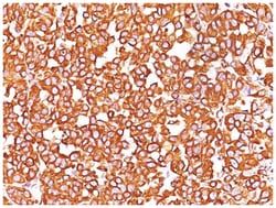
NBP232907B

TYRP1 Mouse, Clone: SPM456, Novus Biologicals™

Manufacturer: Fischer Scientific

The price for this product is unavailable. Please request a quote

Antigen

TYRP1

Classification

Monoclonal

Conjugate

Unconjugated

Formulation

PBS with 0.05% BSA. with 0.05% Sodium Azide

Gene Alias

b-PROTEIN, CAS25,6-dihydroxyindole-2-carboxylic acid oxidase, Catalase B, CATB, EC 1.14.18, EC 1.14.18.-, EC 1.14.18.1, Glycoprotein 75, GP75DHICA oxidase, Melanoma antigen gp75, TRP-1, TRPTYRP, tyrosinase-related protein 1TRP1, TYRRP

Host Species

Mouse

Molecular Weight of Antigen

75 kDa

Quantity

0.2 mg

Primary or Secondary

Primary

Test Specificity

It reacts with a 75kDa melanocyte-specific gene product, identified as Tyrosinase-related protein-1 (TRP-1). It is involved in melanin synthesis. TRP1 is present on the melanosomal membranes of melanoma, normal melanocytes and nevi.Recent evidence suggests that TRP-1 is involved in maintaining stability of tyrosinase protein and modulating its catalytic activity. TRP-1 is also involved in maintenance of melanosome ultrastructure and affects melanocyte proliferation and cell death.

Content And Storage

Store at 4C.

Isotype

IgG2a κ

Applications

Flow Cytometry, Immunocytochemistry, Immunofluorescence, Immunohistochemistry (Frozen)

Clone

SPM456

Dilution

Flow Cytometry 0.5-1ug/million cells, Immunocytochemistry/Immunofluorescence 1-2ug/ml, Immunohistochemistry-Frozen 0.5-1ug/ml

Gene Accession No.

P17643

Gene Symbols

TYRP1

Immunogen

SK-MEL-23 cells

Purification Method

Protein A purified

Regulatory Status

RUO

Gene ID (Entrez)

7306

Target Species

Human, Mouse

Form

Purified

Description

  • TYRP1 Monoclonal specifically detects TYRP1 in Human, Mouse samples
  • It is validated for Flow Cytometry, Immunocytochemistry/Immunofluorescence.

Compare Similar Items

Show Difference

Img

Fischer Scientific

NBP232907B

--


Antigen:
TYRP1

Classification:
Monoclonal

Conjugate:
Unconjugated

Formulation:
PBS with 0.05% BSA. with 0.05% Sodium Azide

Gene Alias:
b-PROTEIN, CAS25,6-dihydroxyindole-2-carboxylic acid oxidase, Catalase B, CATB, EC 1.14.18, EC 1.14.18.-, EC 1.14.18.1, Glycoprotein 75, GP75DHICA oxidase, Melanoma antigen gp75, TRP-1, TRPTYRP, tyrosinase-related protein 1TRP1, TYRRP

Host Species:
Mouse

Molecular Weight of Antigen:
75 kDa

Quantity:
0.2 mg

Primary or Secondary:
Primary

Test Specificity:
It reacts with a 75kDa melanocyte-specific gene product, identified as Tyrosinase-related protein-1 (TRP-1). It is involved in melanin synthesis. TRP1 is present on the melanosomal membranes of melanoma, normal melanocytes and nevi.Recent evidence suggests that TRP-1 is involved in maintaining stability of tyrosinase protein and modulating its catalytic activity. TRP-1 is also involved in maintenance of melanosome ultrastructure and affects melanocyte proliferation and cell death.

Content And Storage:
Store at 4C.

Isotype:
IgG2a κ

Applications:
Flow Cytometry, Immunocytochemistry, Immunofluorescence, Immunohistochemistry (Frozen)

Clone:
SPM456

Dilution:
Flow Cytometry 0.5-1ug/million cells, Immunocytochemistry/Immunofluorescence 1-2ug/ml, Immunohistochemistry-Frozen 0.5-1ug/ml

Gene Accession No.:
P17643

Gene Symbols:
TYRP1

Immunogen:
SK-MEL-23 cells

Purification Method:
Protein A purified

Regulatory Status:
RUO

Gene ID (Entrez):
7306

Target Species:
Human, Mouse

Form:
Purified

Img

Fischer Scientific

NBP232907F

--


Antigen:
TYRP1

Classification:
Monoclonal

Conjugate:
Unconjugated

Formulation:
PBS with 0.05% BSA. with 0.05% Sodium Azide

Gene Alias:
b-PROTEIN, CAS25,6-dihydroxyindole-2-carboxylic acid oxidase, Catalase B, CATB, EC 1.14.18, EC 1.14.18.-, EC 1.14.18.1, Glycoprotein 75, GP75DHICA oxidase, Melanoma antigen gp75, TRP-1, TRPTYRP, tyrosinase-related protein 1TRP1, TYRRP

Host Species:
Mouse

Molecular Weight of Antigen:
75 kDa

Quantity:
0.02 mg

Primary or Secondary:
Primary

Test Specificity:
It reacts with a 75kDa melanocyte-specific gene product, identified as Tyrosinase-related protein-1 (TRP-1). It is involved in melanin synthesis. TRP1 is present on the melanosomal membranes of melanoma, normal melanocytes and nevi.Recent evidence suggests that TRP-1 is involved in maintaining stability of tyrosinase protein and modulating its catalytic activity. TRP-1 is also involved in maintenance of melanosome ultrastructure and affects melanocyte proliferation and cell death.

Content And Storage:
Store at 4C.

Isotype:
IgG2a κ

Applications:
Flow Cytometry, Immunocytochemistry, Immunofluorescence, Immunohistochemistry (Frozen)

Clone:
SPM456

Dilution:
Flow Cytometry 0.5-1ug/million cells, Immunocytochemistry/Immunofluorescence 1-2ug/ml, Immunohistochemistry-Frozen 0.5-1ug/ml

Gene Accession No.:
P17643

Gene Symbols:
TYRP1

Immunogen:
SK-MEL-23 cells

Purification Method:
Protein A purified

Regulatory Status:
RUO

Gene ID (Entrez):
7306

Target Species:
Human, Mouse

Form:
Purified

Img

Fischer Scientific

NBP232910A

--


Antigen:
Vimentin

Classification:
Monoclonal

Conjugate:
Unconjugated

Formulation:
PBS with 0.05% BSA. with 0.05% Sodium Azide

Gene Alias:
FLJ36605, vimentin

Host Species:
Mouse

Molecular Weight of Antigen:
__

Quantity:
0.1 mg

Primary or Secondary:
Primary

Test Specificity:
This MAb reacts with a 58kDa protein identified as vimentin. It shows no cross-reaction with other closely related intermediate filament proteins (IFPs) such as desmin, keratin, neurofilament, and glial fibrillary acid protein.Anti-vimentin alone is of limited value as a diagnostic tool; however, when used in panels with other antibodies, it is useful for the sub-classification of a given tumor. Expression of vimentin, when used in conjunction with anti-keratin, is helpful when distinguishing melanomas from undifferentiated carcinomas and large cell lymphomas. All melanomas and Schwannomas react strongly with anti-vimentin. It labels a variety of mesenchymal cells, including melanocytes, lymphocytes, endothelial cells, and fibroblasts. Non-reactivity of anti-vimentin is often considered more useful than its positive reactivity, since there are a few tumors that do not contain vimentin, e.g. hepatoma and seminoma. Anti-vimentin is also useful as a tissue process control reagent.

Content And Storage:
Store at 4C.

Isotype:
IgG1

Applications:
Western Blot, Flow Cytometry, Immunocytochemistry, Immunofluorescence, Immunoprecipitation, Immunohistochemistry (Paraffin)

Clone:
SPM576

Dilution:
Western Blot 0.5-1ug/ml, Flow Cytometry 0.5-1ug/million cells, Immunocytochemistry/Immunofluorescence 1-2ug/ml, Immunoprecipitation 0.5-1ug/500ug protein lysate, Immunohistochemistry-Paraffin 0.5-1.0ug/ml, Immunohistochemistry-Frozen 0.5-1.0ug/ml

Gene Accession No.:
P08670

Gene Symbols:
VIM

Immunogen:
Human vimentin recombinant protein

Purification Method:
Protein G purified

Regulatory Status:
RUO

Gene ID (Entrez):
7431

Target Species:
Human, Porcine, Bovine, Canine, Chicken, Feline, Goat, Mouse (Negative), Rat (Negative)

Form:
Purified

Img

Fischer Scientific

NBP232910B

--


Antigen:
Vimentin

Classification:
Monoclonal

Conjugate:
Unconjugated

Formulation:
PBS with 0.05% BSA. with 0.05% Sodium Azide

Gene Alias:
FLJ36605, vimentin

Host Species:
Mouse

Molecular Weight of Antigen:
__

Quantity:
0.2 mg

Primary or Secondary:
Primary

Test Specificity:
This MAb reacts with a 58kDa protein identified as vimentin. It shows no cross-reaction with other closely related intermediate filament proteins (IFPs) such as desmin, keratin, neurofilament, and glial fibrillary acid protein.Anti-vimentin alone is of limited value as a diagnostic tool; however, when used in panels with other antibodies, it is useful for the sub-classification of a given tumor. Expression of vimentin, when used in conjunction with anti-keratin, is helpful when distinguishing melanomas from undifferentiated carcinomas and large cell lymphomas. All melanomas and Schwannomas react strongly with anti-vimentin. It labels a variety of mesenchymal cells, including melanocytes, lymphocytes, endothelial cells, and fibroblasts. Non-reactivity of anti-vimentin is often considered more useful than its positive reactivity, since there are a few tumors that do not contain vimentin, e.g. hepatoma and seminoma. Anti-vimentin is also useful as a tissue process control reagent.

Content And Storage:
Store at 4C.

Isotype:
IgG1

Applications:
Western Blot, Flow Cytometry, Immunocytochemistry, Immunofluorescence, Immunoprecipitation, Immunohistochemistry (Paraffin)

Clone:
SPM576

Dilution:
Western Blot 0.5-1ug/ml, Flow Cytometry 0.5-1ug/million cells, Immunocytochemistry/Immunofluorescence 1-2ug/ml, Immunoprecipitation 0.5-1ug/500ug protein lysate, Immunohistochemistry-Paraffin 0.5-1.0ug/ml, Immunohistochemistry-Frozen 0.5-1.0ug/ml

Gene Accession No.:
P08670

Gene Symbols:
VIM

Immunogen:
Human vimentin recombinant protein

Purification Method:
Protein G purified

Regulatory Status:
RUO

Gene ID (Entrez):
7431

Target Species:
Human, Porcine, Bovine, Canine, Chicken, Feline, Goat, Mouse (Negative), Rat (Negative)

Form:
Purified