NBP232925A

CD1b Antibody (RIV12), Novus Biologicals™

Manufacturer: Fischer Scientific

Select a Size

Pack Size SKU Availability Price
Each of 1 NBP232925A-Each-of-1 In Stock ₹ 44,833.44

NBP232925A - Each of 1

₹ 44,833.44

In Stock

Quantity

1

Base Price: ₹ 44,833.44

GST (18%): ₹ 8,070.019

Total Price: ₹ 52,903.459

Antigen

CD1b

Classification

Monoclonal

Conjugate

Unconjugated

Formulation

PBS with 0.05% BSA. with 0.05% Sodium Azide

Gene Alias

CD1, CD1A, CD1b antigen, CD1B antigen, b polypeptide, CD1b molecule, cortical thymocyte antigen CD1B, differentiation antigen CD1-alpha-3, MGC125990, MGC125991, R1, T-cell surface glycoprotein CD1b

Host Species

Mouse

Molecular Weight of Antigen

49 kDa

Quantity

0.1 mg

Research Discipline

Immunology

Gene ID (Entrez)

910

Target Species

Human

Form

Purified

Applications

Flow Cytometry, Immunocytochemistry, Immunofluorescence

Clone

RIV12

Dilution

Flow Cytometry 0.5-1ug/million cells, Immunocytochemistry/Immunofluorescence 0.5-1ug/ml

Gene Accession No.

P29016

Gene Symbols

CD1B

Immunogen

Human peripheral lymphocytes

Purification Method

Protein A purified

Regulatory Status

RUO

Primary or Secondary

Primary

Test Specificity

The mouse monoclonal antibody recognizes CD1b, a 44kDa type I glycoprotein associated with beta2-microglobulin. It is expressed on dendritic cells, Langerhans cells, thymocytes, and T acute lymphoblastic leukemia cells. The CD1 multigene family encodes five forms of the CD1 T-cell surface glycoprotein in human, designated CD1A, 1B, 1C, 1D and 1E. CD1, a type 1 membrane protein, has structural similarity to the MHC class I antigen and has been shown to present lipid antigens for recognition by T lymphocytes. Constitutive endocytosis of CD1B molecules and the differential sorting of MHC class II from lysosomes separate peptide- and lipid antigen-presenting molecules during dendritic cell maturation. CD1B is also expressed in interdigitating cells.

Content And Storage

Store at 4C.

Isotype

IgG1 κ

Description

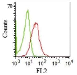
  • CD1b Monoclonal specifically detects CD1b in Human samples
  • It is validated for Flow Cytometry, ELISA, Immunocytochemistry/Immunofluorescence.
Compare Similar Items

Show Difference

Img

Fischer Scientific

NBP232925A

--


Antigen:
CD1b

Classification:
Monoclonal

Conjugate:
Unconjugated

Formulation:
PBS with 0.05% BSA. with 0.05% Sodium Azide

Gene Alias:
CD1, CD1A, CD1b antigen, CD1B antigen, b polypeptide, CD1b molecule, cortical thymocyte antigen CD1B, differentiation antigen CD1-alpha-3, MGC125990, MGC125991, R1, T-cell surface glycoprotein CD1b

Host Species:
Mouse

Molecular Weight of Antigen:
49 kDa

Quantity:
0.1 mg

Research Discipline:
Immunology

Gene ID (Entrez):
910

Target Species:
Human

Form:
Purified

Applications:
Flow Cytometry, Immunocytochemistry, Immunofluorescence

Clone:
RIV12

Dilution:
Flow Cytometry 0.5-1ug/million cells, Immunocytochemistry/Immunofluorescence 0.5-1ug/ml

Gene Accession No.:
P29016

Gene Symbols:
CD1B

Immunogen:
Human peripheral lymphocytes

Purification Method:
Protein A purified

Regulatory Status:
RUO

Primary or Secondary:
Primary

Test Specificity:
The mouse monoclonal antibody recognizes CD1b, a 44kDa type I glycoprotein associated with beta2-microglobulin. It is expressed on dendritic cells, Langerhans cells, thymocytes, and T acute lymphoblastic leukemia cells. The CD1 multigene family encodes five forms of the CD1 T-cell surface glycoprotein in human, designated CD1A, 1B, 1C, 1D and 1E. CD1, a type 1 membrane protein, has structural similarity to the MHC class I antigen and has been shown to present lipid antigens for recognition by T lymphocytes. Constitutive endocytosis of CD1B molecules and the differential sorting of MHC class II from lysosomes separate peptide- and lipid antigen-presenting molecules during dendritic cell maturation. CD1B is also expressed in interdigitating cells.

Content And Storage:
Store at 4C.

Isotype:
IgG1 κ

Img

Fischer Scientific

NBP232925B

--


Antigen:
CD1b

Classification:
Monoclonal

Conjugate:
Unconjugated

Formulation:
PBS with 0.05% BSA. with 0.05% Sodium Azide

Gene Alias:
CD1, CD1A, CD1b antigen, CD1B antigen, b polypeptide, CD1b molecule, cortical thymocyte antigen CD1B, differentiation antigen CD1-alpha-3, MGC125990, MGC125991, R1, T-cell surface glycoprotein CD1b

Host Species:
Mouse

Molecular Weight of Antigen:
49 kDa

Quantity:
0.2 mg

Research Discipline:
Immunology

Gene ID (Entrez):
910

Target Species:
Human

Form:
Purified

Applications:
Flow Cytometry, Immunocytochemistry, Immunofluorescence

Clone:
RIV12

Dilution:
Flow Cytometry 0.5-1ug/million cells, Immunocytochemistry/Immunofluorescence 0.5-1ug/ml

Gene Accession No.:
P29016

Gene Symbols:
CD1B

Immunogen:
Human peripheral lymphocytes

Purification Method:
Protein A purified

Regulatory Status:
RUO

Primary or Secondary:
Primary

Test Specificity:
The mouse monoclonal antibody recognizes CD1b, a 44kDa type I glycoprotein associated with beta2-microglobulin. It is expressed on dendritic cells, Langerhans cells, thymocytes, and T acute lymphoblastic leukemia cells. The CD1 multigene family encodes five forms of the CD1 T-cell surface glycoprotein in human, designated CD1A, 1B, 1C, 1D and 1E. CD1, a type 1 membrane protein, has structural similarity to the MHC class I antigen and has been shown to present lipid antigens for recognition by T lymphocytes. Constitutive endocytosis of CD1B molecules and the differential sorting of MHC class II from lysosomes separate peptide- and lipid antigen-presenting molecules during dendritic cell maturation. CD1B is also expressed in interdigitating cells.

Content And Storage:
Store at 4C.

Isotype:
IgG1 κ

Img

Fischer Scientific

NBP232925F

--


Antigen:
CD1b

Classification:
Monoclonal

Conjugate:
Unconjugated

Formulation:
PBS with 0.05% BSA. with 0.05% Sodium Azide

Gene Alias:
CD1, CD1A, CD1b antigen, CD1B antigen, b polypeptide, CD1b molecule, cortical thymocyte antigen CD1B, differentiation antigen CD1-alpha-3, MGC125990, MGC125991, R1, T-cell surface glycoprotein CD1b

Host Species:
Mouse

Molecular Weight of Antigen:
49 kDa

Quantity:
0.02 mg

Research Discipline:
Immunology

Gene ID (Entrez):
910

Target Species:
Human

Form:
Purified

Applications:
Flow Cytometry, Immunocytochemistry, Immunofluorescence

Clone:
RIV12

Dilution:
Flow Cytometry 0.5-1ug/million cells, Immunocytochemistry/Immunofluorescence 0.5-1ug/ml

Gene Accession No.:
P29016

Gene Symbols:
CD1B

Immunogen:
Human peripheral lymphocytes

Purification Method:
Protein A purified

Regulatory Status:
RUO

Primary or Secondary:
Primary

Test Specificity:
The mouse monoclonal antibody recognizes CD1b, a 44kDa type I glycoprotein associated with beta2-microglobulin. It is expressed on dendritic cells, Langerhans cells, thymocytes, and T acute lymphoblastic leukemia cells. The CD1 multigene family encodes five forms of the CD1 T-cell surface glycoprotein in human, designated CD1A, 1B, 1C, 1D and 1E. CD1, a type 1 membrane protein, has structural similarity to the MHC class I antigen and has been shown to present lipid antigens for recognition by T lymphocytes. Constitutive endocytosis of CD1B molecules and the differential sorting of MHC class II from lysosomes separate peptide- and lipid antigen-presenting molecules during dendritic cell maturation. CD1B is also expressed in interdigitating cells.

Content And Storage:
Store at 4C.

Isotype:
IgG1 κ

Img

Fischer Scientific

NBP232926A

--


Antigen:
CD2

Classification:
Monoclonal

Conjugate:
Unconjugated

Formulation:
PBS with 0.05% BSA. with 0.05% Sodium Azide

Gene Alias:
CD2 antigen, CD2 antigen (p50), sheep red blood cell receptor, CD2 molecule, Erythrocyte receptor, FLJ46032, LFA-2, LFA-3 receptor, lymphocyte-function antigen-2, Rosette receptor, SRBC, T11, T-cell surface antigen CD2, T-cell surface antigen T11/Leu-5

Host Species:
Mouse

Molecular Weight of Antigen:
50 kDa

Quantity:
0.1 mg

Research Discipline:
Adaptive Immunity, Apoptosis, Immunology

Gene ID (Entrez):
914

Target Species:
Human

Form:
Purified

Applications:
Flow Cytometry, Immunocytochemistry, Immunofluorescence, Immunoprecipitation, Immunohistochemistry (Frozen)

Clone:
BH1

Dilution:
Flow Cytometry 0.5-1ug/million cells, Immunocytochemistry/Immunofluorescence 0.5-1ug/ml, Immunoprecipitation 0.5-1ug/500ug protein lysate, Immunohistochemistry-Frozen 0.5-1.0ug/ml

Gene Accession No.:
P06729

Gene Symbols:
CD2

Immunogen:
Human CD2 protein

Purification Method:
Protein A purified

Regulatory Status:
RUO

Primary or Secondary:
Primary

Test Specificity:
CD2 interacts through its amino-terminal domain with the extracellular domain of CD58 (also designated CD2 ligand) to mediate cell adhesion. CD2/CD58 binding can enhance antigen-specific T cell activation. CD2 is a transmembrane glycoprotein that is expressed on peripheral blood T lymphocytes, NK cells and thymocytes. CD58 is a heavily glycosylated protein with a broad tissue distribution in hematopoietic and other cells, including endothelium. Interaction between CD2 and its counter receptor LFA3 (CD58) on opposing cells optimizes immune system recognition, thereby facilitating communication between helper T lymphocytes and antigen-presenting cells, as well as between cytolytic effectors and target cells.

Content And Storage:
Store at 4C.

Isotype:
IgG2b κ