Von Willebrand Factor Antibody (SPM577), Novus Biologicals™
Manufacturer: Fischer Scientific
Select a Size
| Pack Size | SKU | Availability | Price |
|---|---|---|---|
| Each of 1 | NBP233004F-Each-of-1 | In Stock | ₹ 23,852.00 |
NBP233004F - Each of 1
In Stock
Quantity
1
Base Price: ₹ 23,852.00
GST (18%): ₹ 4,293.36
Total Price: ₹ 28,145.36
Antigen
Von Willebrand Factor
Classification
Monoclonal
Conjugate
Unconjugated
Formulation
PBS with 0.05% BSA. with 0.05% Sodium Azide
Gene Alias
coagulation factor VIII VWF, F8, F8VWF, von Willebrand factor, VWD, vWF
Host Species
Mouse
Molecular Weight of Antigen
250 kDa
Quantity
0.02 mg
Research Discipline
Cancer
Gene ID (Entrez)
7450
Target Species
Human
Form
Purified
Applications
Western Blot, Flow Cytometry, Immunocytochemistry, Immunofluorescence, Immunoprecipitation, Immunohistochemistry (Paraffin)
Clone
SPM577
Dilution
Western Blot 0.5-1.0ug/ml, Flow Cytometry 0.5-1ug/million cells, Immunocytochemistry/Immunofluorescence 0.5-1ug/ml, Immunoprecipitation 0.5-1ug/500ug protein lysate, Immunohistochemistry-Paraffin 0.5-1.0ug/ml, Immunohistochemistry-Frozen 0.5-1.0ug/ml
Gene Accession No.
P04275
Gene Symbols
VWF
Immunogen
Recombinant human vWF fragment
Purification Method
Protein A purified
Regulatory Status
RUO
Primary or Secondary
Primary
Test Specificity
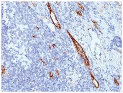
Von Willebrand Factor (vWF) is a multimeric glycoprotein that is found in endothelial cells, plasma and platelets. It acts as a carrier protein for Factor VIII and promotes platelet adhesion and aggregation. vWF undergoes a variety of posttranslational modifications that influence the affinity and availability for Factor VIII, including cleavage of the propeptide and formation of N-terminal disulfide bonds. This antibody helps to establish the endothelial nature of some lesions of disputed histogenesis, e.g. Kaposis sarcoma and cardiac myxoma. It is widely used for differentiating vascular lesions from those of other tissue differentiation within a panel of other vascular markers although not all tumors of endothelial differentiation contain this antigen.
Content And Storage
Store at 4C.
Isotype
IgG1 κ
Description
- Von Willebrand Factor Monoclonal specifically detects Von Willebrand Factor in Human samples
- It is validated for Western Blot, Flow Cytometry, Immunohistochemistry, Immunocytochemistry/Immunofluorescence, Immunohistochemistry-Paraffin.
Compare Similar Items
Show Difference
Antigen: Von Willebrand Factor
Classification: Monoclonal
Conjugate: Unconjugated
Formulation: PBS with 0.05% BSA. with 0.05% Sodium Azide
Gene Alias: coagulation factor VIII VWF, F8, F8VWF, von Willebrand factor, VWD, vWF
Host Species: Mouse
Molecular Weight of Antigen: 250 kDa
Quantity: 0.02 mg
Research Discipline: Cancer
Gene ID (Entrez): 7450
Target Species: Human
Form: Purified
Applications: Western Blot, Flow Cytometry, Immunocytochemistry, Immunofluorescence, Immunoprecipitation, Immunohistochemistry (Paraffin)
Clone: SPM577
Dilution: Western Blot 0.5-1.0ug/ml, Flow Cytometry 0.5-1ug/million cells, Immunocytochemistry/Immunofluorescence 0.5-1ug/ml, Immunoprecipitation 0.5-1ug/500ug protein lysate, Immunohistochemistry-Paraffin 0.5-1.0ug/ml, Immunohistochemistry-Frozen 0.5-1.0ug/ml
Gene Accession No.: P04275
Gene Symbols: VWF
Immunogen: Recombinant human vWF fragment
Purification Method: Protein A purified
Regulatory Status: RUO
Primary or Secondary: Primary
Test Specificity: Von Willebrand Factor (vWF) is a multimeric glycoprotein that is found in endothelial cells, plasma and platelets. It acts as a carrier protein for Factor VIII and promotes platelet adhesion and aggregation. vWF undergoes a variety of posttranslational modifications that influence the affinity and availability for Factor VIII, including cleavage of the propeptide and formation of N-terminal disulfide bonds. This antibody helps to establish the endothelial nature of some lesions of disputed histogenesis, e.g. Kaposis sarcoma and cardiac myxoma. It is widely used for differentiating vascular lesions from those of other tissue differentiation within a panel of other vascular markers although not all tumors of endothelial differentiation contain this antigen.
Content And Storage: Store at 4C.
Isotype: IgG1 κ
Antigen: CEACAM5/CD66e
Classification: Monoclonal
Conjugate: Unconjugated
Formulation: PBS with 0.05% BSA. with 0.05% Sodium Azide
Gene Alias: Carcinoembryonic antigen, carcinoembryonic antigen-related cell adhesion molecule 5, CD66e antigen, CEACD66e, DKFZp781M2392, Meconium antigen 100
Host Species: Mouse
Molecular Weight of Antigen: __
Quantity: 0.1 mg
Research Discipline: __
Gene ID (Entrez): 1048
Target Species: Human, Primate
Form: Purified
Applications: Western Blot, Flow Cytometry, Immunocytochemistry, Immunofluorescence, Immunoprecipitation, Immunohistochemistry (Paraffin)
Clone: C66/261
Dilution: Western Blot 0.25-0.5ug/ml, Flow Cytometry 0.5-1ug/million cells, Immunocytochemistry/Immunofluorescence 1-2ug/ml, Immunoprecipitation 1-2ug/ml, Immunohistochemistry-Paraffin 0.5-1.0ug/ml, Immunohistochemistry-Frozen 0.5-1.0ug/ml
Gene Accession No.: P06731
Gene Symbols: CEACAM5
Immunogen: Recombinant full-length human CEA protein
Purification Method: Protein A purified
Regulatory Status: RUO
Primary or Secondary: Primary
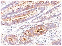
Test Specificity: This antibody recognizes proteins of 80-200kDa, identified as different members of CEA family. CEA is synthesized during development in the fetal gut and is re-expressed in increased amounts in intestinal carcinomas and several other tumors. This MAb does not react with nonspecific cross-reacting antigen (NCA) and with human polymorphonuclear leucocytes. It shows no reaction with a variety of normal tissues and is suitable for staining of formalin/paraffin tissues. CEA is not found in benign glands, stroma, or malignant prostatic cells. Antibody to CEA is useful in detecting early foci of gastric carcinoma and in distinguishing pulmonary adenocarcinomas (60-70% are CEA+) from pleural mesotheliomas (rarely or weakly CEA+). Anti-CEA positivity is seen in adenocarcinomas from the lung, colon, stomach, esophagus, pancreas, gallbadder, urachus, salivary gland, ovary, and endocervix.
Content And Storage: Store at 4C.
Isotype: IgG1 κ
Antigen: CEACAM5/CD66e
Classification: Monoclonal
Conjugate: Unconjugated
Formulation: PBS with 0.05% BSA. with 0.05% Sodium Azide
Gene Alias: Carcinoembryonic antigen, carcinoembryonic antigen-related cell adhesion molecule 5, CD66e antigen, CEACD66e, DKFZp781M2392, Meconium antigen 100
Host Species: Mouse
Molecular Weight of Antigen: __
Quantity: 0.2 mg
Research Discipline: __
Gene ID (Entrez): 1048
Target Species: Human, Primate
Form: Purified
Applications: Western Blot, Flow Cytometry, Immunocytochemistry, Immunofluorescence, Immunoprecipitation, Immunohistochemistry (Paraffin)
Clone: C66/261
Dilution: Western Blot 0.25-0.5ug/ml, Flow Cytometry 0.5-1ug/million cells, Immunocytochemistry/Immunofluorescence 1-2ug/ml, Immunoprecipitation 1-2ug/ml, Immunohistochemistry-Paraffin 0.5-1.0ug/ml, Immunohistochemistry-Frozen 0.5-1.0ug/ml
Gene Accession No.: P06731
Gene Symbols: CEACAM5
Immunogen: Recombinant full-length human CEA protein
Purification Method: Protein A purified
Regulatory Status: RUO
Primary or Secondary: Primary
Test Specificity: This antibody recognizes proteins of 80-200kDa, identified as different members of CEA family. CEA is synthesized during development in the fetal gut and is re-expressed in increased amounts in intestinal carcinomas and several other tumors. This MAb does not react with nonspecific cross-reacting antigen (NCA) and with human polymorphonuclear leucocytes. It shows no reaction with a variety of normal tissues and is suitable for staining of formalin/paraffin tissues. CEA is not found in benign glands, stroma, or malignant prostatic cells. Antibody to CEA is useful in detecting early foci of gastric carcinoma and in distinguishing pulmonary adenocarcinomas (60-70% are CEA+) from pleural mesotheliomas (rarely or weakly CEA+). Anti-CEA positivity is seen in adenocarcinomas from the lung, colon, stomach, esophagus, pancreas, gallbadder, urachus, salivary gland, ovary, and endocervix.
Content And Storage: Store at 4C.
Isotype: IgG1 κ