EpCAM/TROP1 Antibody (VU-1D9), Novus Biologicals™
Manufacturer: Fischer Scientific
Select a Size
| Pack Size | SKU | Availability | Price |
|---|---|---|---|
| Each of 1 | NBP23305102-Each-of-1 | In Stock | ₹ 23,941.00 |
NBP23305102 - Each of 1
In Stock
Quantity
1
Base Price: ₹ 23,941.00
GST (18%): ₹ 4,309.38
Total Price: ₹ 28,250.38
Antigen
EpCAM/TROP1
Classification
Monoclonal
Concentration
0.2mg/mL
Dilution
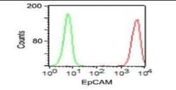
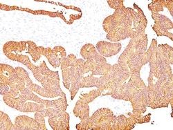
Western Blot 0.5-1ug/ml, Flow Cytometry 0.5-1ug/million cells, ELISA 1-5ug/ml for coating, Immunocytochemistry/Immunofluorescence 1-2ug/ml, Immunoprecipitation 1-2ug/500ug protein, Immunohistochemistry-Paraffin 0.5-1ug/ml, Immunohistochemistry-Frozen 0.5-1ug/ml
Gene Accession No.
P16422
Gene Symbols
EPCAM
Immunogen
Small cell lung carcinoma cells
Quantity
0.02 mg
Research Discipline
Cancer
Gene ID (Entrez)
4072
Target Species
Human, Ferret (Negative), Rat (Negative)
Form
Purified
Applications
Western Blot, Flow Cytometry, ELISA, Immunocytochemistry, Immunofluorescence, Immunoprecipitation, Immunohistochemistry (Paraffin)
Clone
VU-1D9
Conjugate
Unconjugated
Formulation
10mM PBS and 0.05% BSA with 0.05% Sodium Azide
Gene Alias
17-1A, 323/A3, ACSTD1, antigen identified by monoclonal AUA1, CD326 antigen, Cell surface glycoprotein Trop-1, chromosome 4, surface marker (35kD glycoprotein), DIAR5, EGP, EGP-2, EGP314, EGP40, EpCAM, epithelial cell adhesion molecule, Epithelial cell surface antigen, Epithelial glycoprotein, Epithelial glycoprotein 314, ESA, GA733-2EGP34, hEGP314, HNPCC8, KS 1/4 antigen, KS1/4, KSAHEA125, M1S2, M4S1Ly74, Major gastrointestinal tumor-associated protein GA733-2, MIC18MH99, MOC31, TACST-1, TACSTD1, TROP1CD326, Tumor-associated calcium signal transducer 1CO-17A
Host Species
Mouse
Purification Method
Protein G purified
Regulatory Status
RUO
Primary or Secondary
Primary
Test Specificity
This antibody reacts with the first EGF repeat in the extracellular domain of Ep-CAM. It is a 40-43kDa transmembrane epithelial glycoprotein, also identified as epithelial specific antigen (ESA), or epithelial cellular adhesion molecule (Ep-CAM). It is expressed on baso-lateral cell surface in most simple epithelia and a vast majority of carcinomas with the exception of adult squamous epithelium, hepatocytes and gastric epithelial cells. This antibody has been used to distinguish adenocarcinoma from pleural mesothelioma and hepatocellular carcinoma. This antibody is also useful in distinguishing serous carcinomas of the ovary from mesothelioma.
Content And Storage
Store at 4C.
Isotype
IgG1 κ
Related Products


Description
- EpCAM/TROP1 Monoclonal specifically detects EpCAM/TROP1 in Human, Ferret (Negative), Rat (Negative) samples
- It is validated for Western Blot, Flow Cytometry, Immunohistochemistry, Immunocytochemistry/Immunofluorescence, Immunohistochemistry-Paraffin.
Compare Similar Items
Show Difference
Antigen: EpCAM/TROP1
Classification: Monoclonal
Concentration: 0.2mg/mL
Dilution: Western Blot 0.5-1ug/ml, Flow Cytometry 0.5-1ug/million cells, ELISA 1-5ug/ml for coating, Immunocytochemistry/Immunofluorescence 1-2ug/ml, Immunoprecipitation 1-2ug/500ug protein, Immunohistochemistry-Paraffin 0.5-1ug/ml, Immunohistochemistry-Frozen 0.5-1ug/ml
Gene Accession No.: P16422
Gene Symbols: EPCAM
Immunogen: Small cell lung carcinoma cells
Quantity: 0.02 mg
Research Discipline: Cancer
Gene ID (Entrez): 4072
Target Species: Human, Ferret (Negative), Rat (Negative)
Form: Purified
Applications: Western Blot, Flow Cytometry, ELISA, Immunocytochemistry, Immunofluorescence, Immunoprecipitation, Immunohistochemistry (Paraffin)
Clone: VU-1D9
Conjugate: Unconjugated
Formulation: 10mM PBS and 0.05% BSA with 0.05% Sodium Azide
Gene Alias: 17-1A, 323/A3, ACSTD1, antigen identified by monoclonal AUA1, CD326 antigen, Cell surface glycoprotein Trop-1, chromosome 4, surface marker (35kD glycoprotein), DIAR5, EGP, EGP-2, EGP314, EGP40, EpCAM, epithelial cell adhesion molecule, Epithelial cell surface antigen, Epithelial glycoprotein, Epithelial glycoprotein 314, ESA, GA733-2EGP34, hEGP314, HNPCC8, KS 1/4 antigen, KS1/4, KSAHEA125, M1S2, M4S1Ly74, Major gastrointestinal tumor-associated protein GA733-2, MIC18MH99, MOC31, TACST-1, TACSTD1, TROP1CD326, Tumor-associated calcium signal transducer 1CO-17A
Host Species: Mouse
Purification Method: Protein G purified
Regulatory Status: RUO
Primary or Secondary: Primary
Test Specificity: This antibody reacts with the first EGF repeat in the extracellular domain of Ep-CAM. It is a 40-43kDa transmembrane epithelial glycoprotein, also identified as epithelial specific antigen (ESA), or epithelial cellular adhesion molecule (Ep-CAM). It is expressed on baso-lateral cell surface in most simple epithelia and a vast majority of carcinomas with the exception of adult squamous epithelium, hepatocytes and gastric epithelial cells. This antibody has been used to distinguish adenocarcinoma from pleural mesothelioma and hepatocellular carcinoma. This antibody is also useful in distinguishing serous carcinomas of the ovary from mesothelioma.
Content And Storage: Store at 4C.
Isotype: IgG1 κ
Antigen: EpCAM/TROP1
Classification: Monoclonal
Concentration: 0.2mg/mL
Dilution: Western Blot 0.5-1ug/ml, Flow Cytometry 0.5-1ug/million cells, ELISA 1-5ug/ml for coating, Immunocytochemistry/Immunofluorescence 1-2ug/ml, Immunoprecipitation 1-2ug/500ug protein, Immunohistochemistry-Paraffin 0.5-1ug/ml, Immunohistochemistry-Frozen 0.5-1ug/ml
Gene Accession No.: P16422
Gene Symbols: EPCAM
Immunogen: Small cell lung carcinoma cells
Quantity: 0.2 mg
Research Discipline: Cancer
Gene ID (Entrez): 4072
Target Species: Human, Ferret (Negative), Rat (Negative)
Form: Purified
Applications: Western Blot, Flow Cytometry, ELISA, Immunocytochemistry, Immunofluorescence, Immunoprecipitation, Immunohistochemistry (Paraffin)
Clone: VU-1D9
Conjugate: Unconjugated
Formulation: 10mM PBS and 0.05% BSA with 0.05% Sodium Azide
Gene Alias: 17-1A, 323/A3, ACSTD1, antigen identified by monoclonal AUA1, CD326 antigen, Cell surface glycoprotein Trop-1, chromosome 4, surface marker (35kD glycoprotein), DIAR5, EGP, EGP-2, EGP314, EGP40, EpCAM, epithelial cell adhesion molecule, Epithelial cell surface antigen, Epithelial glycoprotein, Epithelial glycoprotein 314, ESA, GA733-2EGP34, hEGP314, HNPCC8, KS 1/4 antigen, KS1/4, KSAHEA125, M1S2, M4S1Ly74, Major gastrointestinal tumor-associated protein GA733-2, MIC18MH99, MOC31, TACST-1, TACSTD1, TROP1CD326, Tumor-associated calcium signal transducer 1CO-17A
Host Species: Mouse
Purification Method: Protein G purified
Regulatory Status: RUO
Primary or Secondary: Primary
Test Specificity: This antibody reacts with the first EGF repeat in the extracellular domain of Ep-CAM. It is a 40-43kDa transmembrane epithelial glycoprotein, also identified as epithelial specific antigen (ESA), or epithelial cellular adhesion molecule (Ep-CAM). It is expressed on baso-lateral cell surface in most simple epithelia and a vast majority of carcinomas with the exception of adult squamous epithelium, hepatocytes and gastric epithelial cells. This antibody has been used to distinguish adenocarcinoma from pleural mesothelioma and hepatocellular carcinoma. This antibody is also useful in distinguishing serous carcinomas of the ovary from mesothelioma.
Content And Storage: Store at 4C.
Isotype: IgG1 κ
Antigen: __
Classification: __
Concentration: 100 mg/mL
Dilution: __
Gene Accession No.: __
Gene Symbols: __
Immunogen: __
Quantity: 0.5 g
Research Discipline: __
Gene ID (Entrez): __
Target Species: __
Form: __
Applications: __
Clone: __
Conjugate: __
Formulation: __
Gene Alias: __
Host Species: __
Purification Method: __
Regulatory Status: __
Primary or Secondary: __
Test Specificity: __
Content And Storage: Store at 4°C short term. Aliquot and store at -20°C long term. Avoid freeze-thaw cycles.
Isotype: __
