Tyrosinase Antibody (T311), Alexa Fluor™ 488, Novus Biologicals™
Manufacturer: Novus Biologicals
Select a Size
| Pack Size | SKU | Availability | Price |
|---|---|---|---|
| Each of 1 | NBP233160X-Each-of-1 | In Stock | ₹ 57,494.00 |
NBP233160X - Each of 1
In Stock
Quantity
1
Base Price: ₹ 57,494.00
GST (18%): ₹ 10,348.92
Total Price: ₹ 67,842.92
Antigen
Tyrosinase
Classification
Monoclonal
Conjugate
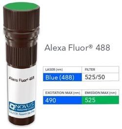
Alexa Fluor 488
Gene Alias
CMM8, EC 1.14.18.1, LB24-AB, Monophenol monooxygenase, OCA1A, OCAIA, SHEP3, SK29-AB, Tumor rejection antigen AB, tyrosinase, tyrosinase (oculocutaneous albinism IA)
Host Species
Mouse
Purification Method
Protein A or G purified
Regulatory Status
RUO
Primary or Secondary
Primary
Test Specificity
Recognizes a cluster of proteins between 70-80kDa, identified as tyrosinase. Occasionally a minor band at 55kDa is also detected. This monoclonal antibody shows no cross-reaction with MAGE-1 and tyrosinase-related protein 1, TRP-1/gp75. Tyrosinase is a copper-containing metalloglycoprotein that catalyzes several steps in the melanin pigment biosynthetic pathway; the hydroxylation of tyrosine to L-3,4-dihydroxy-phenylalanine (dopa), and the subsequent oxidation of dopa to dopaquinone. Mutations of the tyrosinase gene occur in various forms of albinism. Tyrosinase is one of the targets for cytotoxic T-cell recognition in melanoma patients. Staining of melanomas with this monoclonal antibody shows tyrosinase in melanotic as well as amelanotic variants. This monoclonal antibody is a useful marker for melanocytes and melanomas.
Content And Storage
Store at 4C in the dark.
Isotype
IgG2a κ
Applications
Flow Cytometry, Immunohistochemistry, Immunocytochemistry, Immunofluorescence, Immunohistochemistry (Paraffin)
Clone
T311
Dilution
Flow Cytometry, Immunohistochemistry, Immunocytochemistry/Immunofluorescence, Immunohistochemistry-Paraffin
Gene Symbols
TYR
Immunogen
Recombinant tyrosinase protein (Uniprot: P14679 )
Quantity
0.1 mL
Research Discipline
Lipid and Metabolism
Gene ID (Entrez)
7299
Target Species
Human
Form
Purified
Related Products


Description
- Tyrosinase Monoclonal specifically detects Tyrosinase in Human samples
- It is validated for Flow Cytometry, Immunohistochemistry, Immunocytochemistry/Immunofluorescence, Immunohistochemistry-Paraffin.
Compare Similar Items
Show Difference
Antigen: Tyrosinase
Classification: Monoclonal
Conjugate: Alexa Fluor 488
Gene Alias: CMM8, EC 1.14.18.1, LB24-AB, Monophenol monooxygenase, OCA1A, OCAIA, SHEP3, SK29-AB, Tumor rejection antigen AB, tyrosinase, tyrosinase (oculocutaneous albinism IA)
Host Species: Mouse
Purification Method: Protein A or G purified
Regulatory Status: RUO
Primary or Secondary: Primary
Test Specificity: Recognizes a cluster of proteins between 70-80kDa, identified as tyrosinase. Occasionally a minor band at 55kDa is also detected. This monoclonal antibody shows no cross-reaction with MAGE-1 and tyrosinase-related protein 1, TRP-1/gp75. Tyrosinase is a copper-containing metalloglycoprotein that catalyzes several steps in the melanin pigment biosynthetic pathway; the hydroxylation of tyrosine to L-3,4-dihydroxy-phenylalanine (dopa), and the subsequent oxidation of dopa to dopaquinone. Mutations of the tyrosinase gene occur in various forms of albinism. Tyrosinase is one of the targets for cytotoxic T-cell recognition in melanoma patients. Staining of melanomas with this monoclonal antibody shows tyrosinase in melanotic as well as amelanotic variants. This monoclonal antibody is a useful marker for melanocytes and melanomas.
Content And Storage: Store at 4C in the dark.
Isotype: IgG2a κ
Applications: Flow Cytometry, Immunohistochemistry, Immunocytochemistry, Immunofluorescence, Immunohistochemistry (Paraffin)
Clone: T311
Dilution: Flow Cytometry, Immunohistochemistry, Immunocytochemistry/Immunofluorescence, Immunohistochemistry-Paraffin
Gene Symbols: TYR
Immunogen: Recombinant tyrosinase protein (Uniprot: P14679 )
Quantity: 0.1 mL
Research Discipline: Lipid and Metabolism
Gene ID (Entrez): 7299
Target Species: Human
Form: Purified
Antigen: Plasma Cell
Classification: Monoclonal
Conjugate: Biotin
Gene Alias: Plasma
Host Species: Mouse
Purification Method: Protein A or G purified
Regulatory Status: RUO
Primary or Secondary: Primary
Test Specificity: It recognizes an intra-cytoplasmic antigen, which shows a very high degree of specificity for plasma cells. This antigen is present in normal as well as neoplastic plasma cells. Plasma cells, which are large lymphocytes derived from an antigen-specific B cell, secrete antibodies and are responsible for humoral immunity. Plasma cells differentiate from B cells upon stimulation by CD4+ lymphocytes. The B cell acts as an antigen-presenting cell (APC), consuming an offending pathogen, which is taken up by the B cell by phagocytosis and broken down within proteosomes. Plasma cells contain basophilic cytoplasm; their nucleus contains heterochromatin organized in a characteristic cartwheel arrangement. This monoclonal antibody superbly recognizes normal and neoplastic plasma cells in routine formalin-fixed, paraffin-embedded tissue sections. It is of potential value in identifying myeloma or plasmacytoma in bone marrow or other tissues. It also helps differentiate lympho-plasmacytoid lymphoma
Content And Storage: Store at 4C in the dark.
Isotype: IgG2a κ
Applications: Immunohistochemistry (Paraffin)
Clone: LIV3G11 (7B18)
Dilution: Immunohistochemistry-Paraffin
Gene Symbols: __
Immunogen: Pancreatic cancer related serum mucin was used as the immunogen for the Plasma Cell antibody.
Quantity: 0.1 mL
Research Discipline: Immunology
Gene ID (Entrez): __
Target Species: Human, Rat (Negative)
Form: Purified
Antigen: Plasma Cell
Classification: Monoclonal
Conjugate: DyLight 680
Gene Alias: Plasma
Host Species: Mouse
Purification Method: Protein A or G purified
Regulatory Status: RUO
Primary or Secondary: Primary
Test Specificity: It recognizes an intra-cytoplasmic antigen, which shows a very high degree of specificity for plasma cells. This antigen is present in normal as well as neoplastic plasma cells. Plasma cells, which are large lymphocytes derived from an antigen-specific B cell, secrete antibodies and are responsible for humoral immunity. Plasma cells differentiate from B cells upon stimulation by CD4+ lymphocytes. The B cell acts as an antigen-presenting cell (APC), consuming an offending pathogen, which is taken up by the B cell by phagocytosis and broken down within proteosomes. Plasma cells contain basophilic cytoplasm; their nucleus contains heterochromatin organized in a characteristic cartwheel arrangement. This monoclonal antibody superbly recognizes normal and neoplastic plasma cells in routine formalin-fixed, paraffin-embedded tissue sections. It is of potential value in identifying myeloma or plasmacytoma in bone marrow or other tissues. It also helps differentiate lympho-plasmacytoid lymphoma
Content And Storage: Store at 4C in the dark.
Isotype: IgG2a κ
Applications: Immunohistochemistry (Paraffin)
Clone: LIV3G11 (7B18)
Dilution: Immunohistochemistry-Paraffin
Gene Symbols: __
Immunogen: Pancreatic cancer related serum mucin was used as the immunogen for the Plasma Cell antibody.
Quantity: 0.1 mL
Research Discipline: Immunology
Gene ID (Entrez): __
Target Species: Human, Rat (Negative)
Form: Purified