Retinol Binding Protein RBP Mouse, Clone: G4E4, Novus Biologicals™
Manufacturer: Fischer Scientific
Select a Size
| Pack Size | SKU | Availability | Price |
|---|---|---|---|
| Each of 1 | NBP234231A-Each-of-1 | In Stock | ₹ 45,078.50 |
NBP234231A - Each of 1
In Stock
Quantity
1
Base Price: ₹ 45,078.50
GST (18%): ₹ 8,114.13
Total Price: ₹ 53,192.63
Antigen
Retinol Binding Protein RBP
Classification
Monoclonal
Conjugate
Unconjugated
Formulation
PBS with 0.05% BSA. with 0.05% Sodium Azide
Gene Alias
C, Cellular retinol-binding protein, CRABP-I, CRBP1CRBP-I, CRBPCellular retinol-binding protein I, CRBPI, RBPC, retinol binding protein 1, cellular, retinol-binding protein 1, retinol-binding protein 1, cellular
Host Species
Mouse
Molecular Weight of Antigen
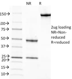
23 kDa
Quantity
0.1 mg
Primary or Secondary
Primary
Test Specificity
This MAb recognizes an epitope within the 74-182 C-terminal sequence (11kD peptide fragment) of human serum Cellular Retinol Binding Protein 1 (CRBP 1), a single-chain glycoprotein belonging to the superfamily of hydrophobic molecule transporter proteins, which is responsible for transport of retinol (vitamin A1) from the liver to peripheral target tissues, like the eye, where it mediates the cellular uptake. CRBP 1 is synthesized by hepatic parenchymal cells where it becomes bound to its ligand retinol and is then released into the circulation, where it binds further to the protein transthyretin, to form a transporting complex, which is big enough not to be lost by filtration through the kidney glomeruli. It is detected in nearly all tissues with higher expression in adult ovary, pancreas, pituitary gland, adrenal gland, and fetal liver.
Content And Storage
Store at 4C.
Isotype
IgG1 κ
Applications
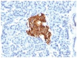
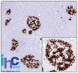
Western Blot, Immunocytochemistry, Immunofluorescence, Immunoprecipitation, Immunohistochemistry (Frozen)
Clone
G4E4
Dilution
Western Blot 0.5-1ug/ml, Immunocytochemistry/Immunofluorescence 1-2ug/ml, Immunoprecipitation 1-2ug/500ug protein, Immunohistochemistry-Frozen 1-2ug/ml, SDS-Page
Gene Accession No.
P02753
Gene Symbols
RBP1
Immunogen
Retinol binding protein-1 purified from human plasma
Purification Method
Protein A purified
Regulatory Status
RUO
Gene ID (Entrez)
5947
Target Species
Human, Mouse, Rat, Goat, Primate, Rabbit
Form
Purified
Related Products


Description
- Retinol Binding Protein RBP Monoclonal specifically detects Retinol Binding Protein RBP in Human, Mouse, Rat, Goat, Chimpanzee, Monkey, Rabbit samples
- It is validated for Immunohistochemistry, Immunohistochemistry-Paraffin.
Compare Similar Items
Show Difference
Antigen: Retinol Binding Protein RBP
Classification: Monoclonal
Conjugate: Unconjugated
Formulation: PBS with 0.05% BSA. with 0.05% Sodium Azide
Gene Alias: C, Cellular retinol-binding protein, CRABP-I, CRBP1CRBP-I, CRBPCellular retinol-binding protein I, CRBPI, RBPC, retinol binding protein 1, cellular, retinol-binding protein 1, retinol-binding protein 1, cellular
Host Species: Mouse
Molecular Weight of Antigen: 23 kDa
Quantity: 0.1 mg
Primary or Secondary: Primary
Test Specificity: This MAb recognizes an epitope within the 74-182 C-terminal sequence (11kD peptide fragment) of human serum Cellular Retinol Binding Protein 1 (CRBP 1), a single-chain glycoprotein belonging to the superfamily of hydrophobic molecule transporter proteins, which is responsible for transport of retinol (vitamin A1) from the liver to peripheral target tissues, like the eye, where it mediates the cellular uptake. CRBP 1 is synthesized by hepatic parenchymal cells where it becomes bound to its ligand retinol and is then released into the circulation, where it binds further to the protein transthyretin, to form a transporting complex, which is big enough not to be lost by filtration through the kidney glomeruli. It is detected in nearly all tissues with higher expression in adult ovary, pancreas, pituitary gland, adrenal gland, and fetal liver.
Content And Storage: Store at 4C.
Isotype: IgG1 κ
Applications: Western Blot, Immunocytochemistry, Immunofluorescence, Immunoprecipitation, Immunohistochemistry (Frozen)
Clone: G4E4
Dilution: Western Blot 0.5-1ug/ml, Immunocytochemistry/Immunofluorescence 1-2ug/ml, Immunoprecipitation 1-2ug/500ug protein, Immunohistochemistry-Frozen 1-2ug/ml, SDS-Page
Gene Accession No.: P02753
Gene Symbols: RBP1
Immunogen: Retinol binding protein-1 purified from human plasma
Purification Method: Protein A purified
Regulatory Status: RUO
Gene ID (Entrez): 5947
Target Species: Human, Mouse, Rat, Goat, Primate, Rabbit
Form: Purified
Antigen: Retinol Binding Protein RBP
Classification: Monoclonal
Conjugate: Unconjugated
Formulation: PBS with 0.05% BSA. with 0.05% Sodium Azide
Gene Alias: C, Cellular retinol-binding protein, CRABP-I, CRBP1CRBP-I, CRBPCellular retinol-binding protein I, CRBPI, RBPC, retinol binding protein 1, cellular, retinol-binding protein 1, retinol-binding protein 1, cellular
Host Species: Mouse
Molecular Weight of Antigen: 23 kDa
Quantity: 0.2 mg
Primary or Secondary: Primary
Test Specificity: This MAb recognizes an epitope within the 74-182 C-terminal sequence (11kD peptide fragment) of human serum Cellular Retinol Binding Protein 1 (CRBP 1), a single-chain glycoprotein belonging to the superfamily of hydrophobic molecule transporter proteins, which is responsible for transport of retinol (vitamin A1) from the liver to peripheral target tissues, like the eye, where it mediates the cellular uptake. CRBP 1 is synthesized by hepatic parenchymal cells where it becomes bound to its ligand retinol and is then released into the circulation, where it binds further to the protein transthyretin, to form a transporting complex, which is big enough not to be lost by filtration through the kidney glomeruli. It is detected in nearly all tissues with higher expression in adult ovary, pancreas, pituitary gland, adrenal gland, and fetal liver.
Content And Storage: Store at 4C.
Isotype: IgG1 κ
Applications: Western Blot, Immunocytochemistry, Immunofluorescence, Immunoprecipitation, Immunohistochemistry (Frozen)
Clone: G4E4
Dilution: Western Blot 0.5-1ug/ml, Immunocytochemistry/Immunofluorescence 1-2ug/ml, Immunoprecipitation 1-2ug/500ug protein, Immunohistochemistry-Frozen 1-2ug/ml, SDS-Page
Gene Accession No.: P02753
Gene Symbols: RBP1
Immunogen: Retinol binding protein-1 purified from human plasma
Purification Method: Protein A purified
Regulatory Status: RUO
Gene ID (Entrez): 5947
Target Species: Human, Mouse, Rat, Goat, Primate, Rabbit
Form: Purified
Antigen: Retinol Binding Protein RBP
Classification: Monoclonal
Conjugate: Unconjugated
Formulation: PBS with 0.05% BSA. with 0.05% Sodium Azide
Gene Alias: C, Cellular retinol-binding protein, CRABP-I, CRBP1CRBP-I, CRBPCellular retinol-binding protein I, CRBPI, RBPC, retinol binding protein 1, cellular, retinol-binding protein 1, retinol-binding protein 1, cellular
Host Species: Mouse
Molecular Weight of Antigen: 23 kDa
Quantity: 0.02 mg
Primary or Secondary: Primary
Test Specificity: This MAb recognizes an epitope within the 74-182 C-terminal sequence (11kD peptide fragment) of human serum Cellular Retinol Binding Protein 1 (CRBP 1), a single-chain glycoprotein belonging to the superfamily of hydrophobic molecule transporter proteins, which is responsible for transport of retinol (vitamin A1) from the liver to peripheral target tissues, like the eye, where it mediates the cellular uptake. CRBP 1 is synthesized by hepatic parenchymal cells where it becomes bound to its ligand retinol and is then released into the circulation, where it binds further to the protein transthyretin, to form a transporting complex, which is big enough not to be lost by filtration through the kidney glomeruli. It is detected in nearly all tissues with higher expression in adult ovary, pancreas, pituitary gland, adrenal gland, and fetal liver.
Content And Storage: Store at 4C.
Isotype: IgG1 κ
Applications: Western Blot, Immunocytochemistry, Immunofluorescence, Immunoprecipitation, Immunohistochemistry (Frozen)
Clone: G4E4
Dilution: Western Blot 0.5-1ug/ml, Immunocytochemistry/Immunofluorescence 1-2ug/ml, Immunoprecipitation 1-2ug/500ug protein, Immunohistochemistry-Frozen 1-2ug/ml, SDS-Page
Gene Accession No.: P02753
Gene Symbols: RBP1
Immunogen: Retinol binding protein-1 purified from human plasma
Purification Method: Protein A purified
Regulatory Status: RUO
Gene ID (Entrez): 5947
Target Species: Human, Mouse, Rat, Goat, Primate, Rabbit
Form: Purified
