Myogenin Mouse, Clone: MGN185 + F5D, Novus Biologicals™
Manufacturer: Fischer Scientific
Select a Size
| Pack Size | SKU | Availability | Price |
|---|---|---|---|
| Each of 1 | NBP234278F-Each-of-1 | In Stock | ₹ 23,852.00 |
NBP234278F - Each of 1
In Stock
Quantity
1
Base Price: ₹ 23,852.00
GST (18%): ₹ 4,293.36
Total Price: ₹ 28,145.36
Antigen
Myogenin
Classification
Monoclonal
Concentration
0.2mg/mL
Dilution
Western Blot 0.5-1.0ug/ml, Flow Cytometry 0.5-1ug/million cells, Immunocytochemistry/Immunofluorescence 0.5-1ug/ml, Immunoprecipitation 0.5-1ug/500ug protein lysate, Immunohistochemistry-Paraffin 0.5-1ug/ml, Immunohistochemistry-Frozen 0.5-1ug/ml
Gene Accession No.
P15173
Gene Symbols
MYOG
Immunogen
Human myogenin recombinant protein (MGN185); Rat myogenin recombinant fragment containing amino acid 30-224 (F5D)
Purification Method
Protein A purified
Regulatory Status
RUO
Primary or Secondary
Primary
Test Specificity
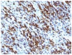
Myogenin is a member of the MyoD family of myogenic basic helix-loop-helix (bHLH) transcription factors that also includes MyoD, Myf-5, and MRF4 (also known as herculinor Myf-6). MyoD family members are expressed exclusively in skeletal muscle and play a key role in activating myogenesis by binding to enhancer sequences of muscle-specific genes. The regulatory domain of MyoD is approximately 70 amino acids in length and includes both a basic DNA binding motif and a bHLH dimerization motif. MyoD family members share about 80% amino acid homology in their bHLH motifs. Anti-myogenin labels the nuclei of myoblasts in developing muscle tissue, and is expressed in tumor cell nuclei of rhabdomyosarcoma and some leiomyosarcomas. Positive nuclear staining may occur in Wilms tumor.
Content And Storage
Store at 4C.
Isotype
IgG
Applications
Western Blot, Flow Cytometry, Immunocytochemistry, Immunofluorescence, Immunoprecipitation, Immunohistochemistry (Paraffin)
Clone
MGN185 + F5D
Conjugate
Unconjugated
Formulation
PBS with 0.05% BSA. with 0.05% Sodium Azide
Gene Alias
BHLHC3, bHLHc3Myogenic factor-4; myogenin, Class C basic helix-loop-helix protein 3, myf-4, MYF4myogenin, Myogenic factor 4, MYOGENIN, myogenin (myogenic factor 4)
Host Species
Mouse
Molecular Weight of Antigen
34 kDa
Quantity
0.02 mg
Research Discipline
Cancer, Stem Cell Markers, Transcription Factors and Regulators
Gene ID (Entrez)
4656
Target Species
Human, Mouse, Rat, Porcine, Feline
Form
Purified
Related Products

Description
- Myogenin Monoclonal specifically detects Myogenin in Human, Mouse, Rat, Porcine, Feline samples
- It is validated for Immunohistochemistry, Immunohistochemistry-Paraffin.
Compare Similar Items
Show Difference
Antigen: Myogenin
Classification: Monoclonal
Concentration: 0.2mg/mL
Dilution: Western Blot 0.5-1.0ug/ml, Flow Cytometry 0.5-1ug/million cells, Immunocytochemistry/Immunofluorescence 0.5-1ug/ml, Immunoprecipitation 0.5-1ug/500ug protein lysate, Immunohistochemistry-Paraffin 0.5-1ug/ml, Immunohistochemistry-Frozen 0.5-1ug/ml
Gene Accession No.: P15173
Gene Symbols: MYOG
Immunogen: Human myogenin recombinant protein (MGN185); Rat myogenin recombinant fragment containing amino acid 30-224 (F5D)
Purification Method: Protein A purified
Regulatory Status: RUO
Primary or Secondary: Primary
Test Specificity: Myogenin is a member of the MyoD family of myogenic basic helix-loop-helix (bHLH) transcription factors that also includes MyoD, Myf-5, and MRF4 (also known as herculinor Myf-6). MyoD family members are expressed exclusively in skeletal muscle and play a key role in activating myogenesis by binding to enhancer sequences of muscle-specific genes. The regulatory domain of MyoD is approximately 70 amino acids in length and includes both a basic DNA binding motif and a bHLH dimerization motif. MyoD family members share about 80% amino acid homology in their bHLH motifs. Anti-myogenin labels the nuclei of myoblasts in developing muscle tissue, and is expressed in tumor cell nuclei of rhabdomyosarcoma and some leiomyosarcomas. Positive nuclear staining may occur in Wilms tumor.
Content And Storage: Store at 4C.
Isotype: IgG
Applications: Western Blot, Flow Cytometry, Immunocytochemistry, Immunofluorescence, Immunoprecipitation, Immunohistochemistry (Paraffin)
Clone: MGN185 + F5D
Conjugate: Unconjugated
Formulation: PBS with 0.05% BSA. with 0.05% Sodium Azide
Gene Alias: BHLHC3, bHLHc3Myogenic factor-4; myogenin, Class C basic helix-loop-helix protein 3, myf-4, MYF4myogenin, Myogenic factor 4, MYOGENIN, myogenin (myogenic factor 4)
Host Species: Mouse
Molecular Weight of Antigen: 34 kDa
Quantity: 0.02 mg
Research Discipline: Cancer, Stem Cell Markers, Transcription Factors and Regulators
Gene ID (Entrez): 4656
Target Species: Human, Mouse, Rat, Porcine, Feline
Form: Purified
Antigen: NCAM-1/CD56
Classification: Monoclonal
Concentration: __
Dilution: Western Blot 0.5-1ug/ml, Flow Cytometry 0.5-1ug/million cells, Immunocytochemistry/Immunofluorescence 1-2ug/ml, Immunoprecipitation 1-2ug/500ug protein lysate, Immunohistochemistry-Paraffin 0.5-1ug/ml, Immunohistochemistry-Frozen 0.5-1ug/ml
Gene Accession No.: P13591
Gene Symbols: NCAM1
Immunogen: Membrane preparation of a small cell lung carcinoma
Purification Method: Protein A purified
Regulatory Status: RUO
Primary or Secondary: Primary
Test Specificity: This MAb reacts with an extracellular domain (close to transmembrane) of CD56/NCAM. Three isoforms of neural cell adhesion molecule (NCAM) are produced by differential splicing of the RNA transcript from a single gene. The 135kDa isoform is the basic molecule, which is glycosylated or sialylated to produce the mature species. Anti-CD56 recognizes two proteins of the neural cell adhesion molecule, the basic molecule expressed on most neuroectodermally derived tissues and neoplasms (e.g. retinoblastoma, medulloblastomas, astrocytomas, neuroblastomas, and small cell carcinomas). It is also expressed on some mesodermally derived tumors (rhabdomyosarcoma). Anti-CD56 plays an important role in the diagnosis of nodal and nasal NK/T-cell lymphomas.
Content And Storage: Store at 4C.
Isotype: IgG1 κ
Applications: Western Blot, Flow Cytometry, Immunocytochemistry, Immunofluorescence, Immunoprecipitation, Immunohistochemistry (Paraffin)
Clone: 123A8 (same as 56C04)
Conjugate: Unconjugated
Formulation: PBS with 0.05% BSA. with 0.05% Sodium Azide
Gene Alias: CD56, CD56/ NCAM-1, CD56 antigen, MSK39, N-CAM-1, NCAM-1, NCAMantigen recognized by monoclonal 5.1H11, neural cell adhesion molecule 1, neural cell adhesion molecule, NCAM
Host Species: Mouse
Molecular Weight of Antigen: __
Quantity: 0.1 mg
Research Discipline: Astrocyte Markers, Cellular Markers, Cytokine Research, Cytoskeleton Markers, Growth and Development, Hematopoietic Stem Cell Markers, Immunology, Innate Immunity, Membrane Vesicle Markers, Neuronal Cell Markers, Neuronal Stem Cell Markers, Neuroscience, Stem Cell Markers
Gene ID (Entrez): 4684
Target Species: Human
Form: Purified
Antigen: NCAM-1/CD56
Classification: Monoclonal
Concentration: __
Dilution: Western Blot 0.5-1ug/ml, Flow Cytometry 0.5-1ug/million cells, Immunocytochemistry/Immunofluorescence 1-2ug/ml, Immunoprecipitation 1-2ug/500ug protein lysate, Immunohistochemistry-Paraffin 0.5-1ug/ml, Immunohistochemistry-Frozen 0.5-1ug/ml
Gene Accession No.: P13591
Gene Symbols: NCAM1
Immunogen: Membrane preparation of a small cell lung carcinoma
Purification Method: Protein A purified
Regulatory Status: RUO
Primary or Secondary: Primary
Test Specificity: This MAb reacts with an extracellular domain (close to transmembrane) of CD56/NCAM. Three isoforms of neural cell adhesion molecule (NCAM) are produced by differential splicing of the RNA transcript from a single gene. The 135kDa isoform is the basic molecule, which is glycosylated or sialylated to produce the mature species. Anti-CD56 recognizes two proteins of the neural cell adhesion molecule, the basic molecule expressed on most neuroectodermally derived tissues and neoplasms (e.g. retinoblastoma, medulloblastomas, astrocytomas, neuroblastomas, and small cell carcinomas). It is also expressed on some mesodermally derived tumors (rhabdomyosarcoma). Anti-CD56 plays an important role in the diagnosis of nodal and nasal NK/T-cell lymphomas.
Content And Storage: Store at 4C.
Isotype: IgG1 κ
Applications: Western Blot, Flow Cytometry, Immunocytochemistry, Immunofluorescence, Immunoprecipitation, Immunohistochemistry (Paraffin)
Clone: 123A8 (same as 56C04)
Conjugate: Unconjugated
Formulation: PBS with 0.05% BSA. with 0.05% Sodium Azide
Gene Alias: CD56, CD56/ NCAM-1, CD56 antigen, MSK39, N-CAM-1, NCAM-1, NCAMantigen recognized by monoclonal 5.1H11, neural cell adhesion molecule 1, neural cell adhesion molecule, NCAM
Host Species: Mouse
Molecular Weight of Antigen: __
Quantity: 0.2 mg
Research Discipline: Astrocyte Markers, Cellular Markers, Cytokine Research, Cytoskeleton Markers, Growth and Development, Hematopoietic Stem Cell Markers, Immunology, Innate Immunity, Membrane Vesicle Markers, Neuronal Cell Markers, Neuronal Stem Cell Markers, Neuroscience, Stem Cell Markers
Gene ID (Entrez): 4684
Target Species: Human
Form: Purified