Mitochondria Antibody (113-1), Biotin, Novus Biologicals™
Manufacturer: Novus Biologicals
Select a Size
| Pack Size | SKU | Availability | Price |
|---|---|---|---|
| Each of 1 | NBP234517B-Each-of-1 | In Stock | ₹ 57,494.00 |
NBP234517B - Each of 1
In Stock
Quantity
1
Base Price: ₹ 57,494.00
GST (18%): ₹ 10,348.92
Total Price: ₹ 67,842.92
Antigen
Mitochondria
Classification
Monoclonal
Conjugate
Biotin
Host Species
Mouse
Molecular Weight of Antigen
60 kDa
Quantity
0.1 mL
Primary or Secondary
Primary
Target Species
Human, Mouse (Negative), Rat (Negative)
Form
Purified
Applications
Western Blot, Immunocytochemistry, Immunofluorescence, Immunohistochemistry (Paraffin), Immunohistochemistry (Frozen)
Clone
113-1
Dilution
Western Blot, Immunocytochemistry/Immunofluorescence, Immunohistochemistry-Paraffin, Immunohistochemistry-Frozen
Immunogen
Semi-purified mitochondrial preparation
Purification Method
Protein A or G purified
Regulatory Status
RUO
Test Specificity
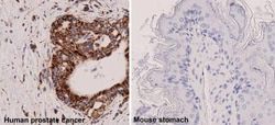
This monoclonal antibody recognizes a 60kDa antigen associated with the mitochondria in human cells. It can be used to stain mitochondria in cell or tissue preparations and can be used as a mitochondrial marker in subcellular fractions. It produces a spaghetti-like pattern in normal and malignant cells. This antibody is an excellent marker for human cells in xenographic model research. It reacts specifically with human cells, including neurons and embryonic stem cells. Immunostaining pattern with anti-mitochondrial monoclonal antibody has been reported as a useful discriminatory adjunct in the complex differential diagnosis of granular renal cell tumors. Reportedly, this monoclonal antibody facilitates the classification of salivary tumors.
Content And Storage
Store at 4C in the dark.
Isotype
IgG1 κ
Related Products

Description
- Mitochondria Monoclonal specifically detects Mitochondria in Human, Mouse (Negative), Rat (Negative) samples
- It is validated for Western Blot, Immunohistochemistry, Immunocytochemistry/Immunofluorescence, Immunohistochemistry-Paraffin, Immunohistochemistry-Frozen.
Compare Similar Items
Show Difference
Antigen: Mitochondria
Classification: Monoclonal
Conjugate: Biotin
Host Species: Mouse
Molecular Weight of Antigen: 60 kDa
Quantity: 0.1 mL
Primary or Secondary: Primary
Target Species: Human, Mouse (Negative), Rat (Negative)
Form: Purified
Applications: Western Blot, Immunocytochemistry, Immunofluorescence, Immunohistochemistry (Paraffin), Immunohistochemistry (Frozen)
Clone: 113-1
Dilution: Western Blot, Immunocytochemistry/Immunofluorescence, Immunohistochemistry-Paraffin, Immunohistochemistry-Frozen
Immunogen: Semi-purified mitochondrial preparation
Purification Method: Protein A or G purified
Regulatory Status: RUO
Test Specificity: This monoclonal antibody recognizes a 60kDa antigen associated with the mitochondria in human cells. It can be used to stain mitochondria in cell or tissue preparations and can be used as a mitochondrial marker in subcellular fractions. It produces a spaghetti-like pattern in normal and malignant cells. This antibody is an excellent marker for human cells in xenographic model research. It reacts specifically with human cells, including neurons and embryonic stem cells. Immunostaining pattern with anti-mitochondrial monoclonal antibody has been reported as a useful discriminatory adjunct in the complex differential diagnosis of granular renal cell tumors. Reportedly, this monoclonal antibody facilitates the classification of salivary tumors.
Content And Storage: Store at 4C in the dark.
Isotype: IgG1 κ
Antigen: Mitochondria
Classification: Monoclonal
Conjugate: DyLight 650
Host Species: Mouse
Molecular Weight of Antigen: 60 kDa
Quantity: 0.1 mL
Primary or Secondary: Primary
Target Species: Human, Mouse (Negative), Rat (Negative)
Form: Purified
Applications: Western Blot, Immunocytochemistry, Immunofluorescence, Immunohistochemistry (Paraffin), Immunohistochemistry (Frozen)
Clone: 113-1
Dilution: Western Blot, Immunocytochemistry/Immunofluorescence, Immunohistochemistry-Paraffin, Immunohistochemistry-Frozen
Immunogen: Semi-purified mitochondrial preparation
Purification Method: Protein A or G purified
Regulatory Status: RUO
Test Specificity: This monoclonal antibody recognizes a 60kDa antigen associated with the mitochondria in human cells. It can be used to stain mitochondria in cell or tissue preparations and can be used as a mitochondrial marker in subcellular fractions. It produces a spaghetti-like pattern in normal and malignant cells. This antibody is an excellent marker for human cells in xenographic model research. It reacts specifically with human cells, including neurons and embryonic stem cells. Immunostaining pattern with anti-mitochondrial monoclonal antibody has been reported as a useful discriminatory adjunct in the complex differential diagnosis of granular renal cell tumors. Reportedly, this monoclonal antibody facilitates the classification of salivary tumors.
Content And Storage: Store at 4C in the dark.
Isotype: IgG1 κ
Antigen: Mitochondria
Classification: Monoclonal
Conjugate: DyLight 350
Host Species: Mouse
Molecular Weight of Antigen: 60 kDa
Quantity: 0.1 mL
Primary or Secondary: Primary
Target Species: Human, Mouse (Negative), Rat (Negative)
Form: Purified
Applications: Western Blot, Immunocytochemistry, Immunofluorescence, Immunohistochemistry (Paraffin), Immunohistochemistry (Frozen)
Clone: 113-1
Dilution: Western Blot, Immunocytochemistry/Immunofluorescence, Immunohistochemistry-Paraffin, Immunohistochemistry-Frozen
Immunogen: Semi-purified mitochondrial preparation
Purification Method: Protein A or G purified
Regulatory Status: RUO
Test Specificity: This monoclonal antibody recognizes a 60kDa antigen associated with the mitochondria in human cells. It can be used to stain mitochondria in cell or tissue preparations and can be used as a mitochondrial marker in subcellular fractions. It produces a spaghetti-like pattern in normal and malignant cells. This antibody is an excellent marker for human cells in xenographic model research. It reacts specifically with human cells, including neurons and embryonic stem cells. Immunostaining pattern with anti-mitochondrial monoclonal antibody has been reported as a useful discriminatory adjunct in the complex differential diagnosis of granular renal cell tumors. Reportedly, this monoclonal antibody facilitates the classification of salivary tumors.
Content And Storage: Store at 4C in the dark.
Isotype: IgG1 κ