NBP234752W

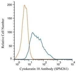
Cytokeratin 10 Antibody (SPM261), Alexa Fluor™ 405, Novus Biologicals™

Manufacturer: Novus Biologicals

Select a Size

Pack Size SKU Availability Price
Each of 1 NBP234752W-Each-of-1 In Stock ₹ 55,271.76

NBP234752W - Each of 1

₹ 55,271.76

In Stock

Quantity

1

Base Price: ₹ 55,271.76

GST (18%): ₹ 9,948.917

Total Price: ₹ 65,220.677

Antigen

Cytokeratin 10

Classification

Monoclonal

Conjugate

Alexa Fluor 405

Gene Alias

BCIE, BIE, CK10, CK-10, cytokeratin 10, Cytokeratin-10, EHK, K10keratosis palmaris et plantaris, keratin 10, Keratin 10 (epidermolytic hyperkeratosis; keratosis palmaris et plantaris), keratin, type I cytoskeletal 10, keratin-10, KPP

Host Species

Mouse

Molecular Weight of Antigen

56.5 kDa

Quantity

0.1 mL

Research Discipline

Cytoskeleton Markers

Gene ID (Entrez)

3858

Target Species

Human, Mouse

Form

Purified

Applications

Western Blot, Flow Cytometry, ELISA, Immunohistochemistry, Immunocytochemistry, Immunofluorescence, Immunohistochemistry (Paraffin), Immunohistochemistry (Frozen)

Clone

SPM261

Dilution

Western Blot, Flow Cytometry, ELISA, Immunohistochemistry, Immunocytochemistry/Immunofluorescence, Immunohistochemistry-Paraffin, Immunohistochemistry-Frozen

Gene Symbols

KRT10

Immunogen

Skin extract of a human Psoriasis patient

Purification Method

Protein A or G purified

Regulatory Status

RUO

Primary or Secondary

Primary

Test Specificity

This MAb recognizes a protein of 56.5kDa, identified as cytokeratin 10 (CK10). CK10 is expressed in all suprabasal layers of the epidermis. In the epidermis, expression of CK10 strictly parallels the extent of differentiation; it is absent in the basal layer, appears in the first suprabasal layers and increases in concentration towards the granular layer. However, CK10 is rarely detected in early stages of vulvar squamous carcinomas (tumors less than 2 cm, clinical stage I) regardless of the tumor grade. In larger and more advanced tumors (greater than 2 cm, clinical stages II and III), CK10 is detected very frequently. Expression of CK10 is related to maturation of malignant keratinocytes, being preferentially detected in more-differentiated parts.

Content And Storage

Store at 4C in the dark.

Isotype

IgG1 κ

Description

  • Cytokeratin 10 Monoclonal specifically detects Cytokeratin 10 in Human, Mouse samples
  • It is validated for Flow Cytometry, Immunohistochemistry, Immunohistochemistry-Paraffin.

Compare Similar Items

Show Difference

Img

Novus Biologicals

NBP234752W

--


Antigen:
Cytokeratin 10

Classification:
Monoclonal

Conjugate:
Alexa Fluor 405

Gene Alias:
BCIE, BIE, CK10, CK-10, cytokeratin 10, Cytokeratin-10, EHK, K10keratosis palmaris et plantaris, keratin 10, Keratin 10 (epidermolytic hyperkeratosis; keratosis palmaris et plantaris), keratin, type I cytoskeletal 10, keratin-10, KPP

Host Species:
Mouse

Molecular Weight of Antigen:
56.5 kDa

Quantity:
0.1 mL

Research Discipline:
Cytoskeleton Markers

Gene ID (Entrez):
3858

Target Species:
Human, Mouse

Form:
Purified

Applications:
Western Blot, Flow Cytometry, ELISA, Immunohistochemistry, Immunocytochemistry, Immunofluorescence, Immunohistochemistry (Paraffin), Immunohistochemistry (Frozen)

Clone:
SPM261

Dilution:
Western Blot, Flow Cytometry, ELISA, Immunohistochemistry, Immunocytochemistry/Immunofluorescence, Immunohistochemistry-Paraffin, Immunohistochemistry-Frozen

Gene Symbols:
KRT10

Immunogen:
Skin extract of a human Psoriasis patient

Purification Method:
Protein A or G purified

Regulatory Status:
RUO

Primary or Secondary:
Primary

Test Specificity:
This MAb recognizes a protein of 56.5kDa, identified as cytokeratin 10 (CK10). CK10 is expressed in all suprabasal layers of the epidermis. In the epidermis, expression of CK10 strictly parallels the extent of differentiation; it is absent in the basal layer, appears in the first suprabasal layers and increases in concentration towards the granular layer. However, CK10 is rarely detected in early stages of vulvar squamous carcinomas (tumors less than 2 cm, clinical stage I) regardless of the tumor grade. In larger and more advanced tumors (greater than 2 cm, clinical stages II and III), CK10 is detected very frequently. Expression of CK10 is related to maturation of malignant keratinocytes, being preferentially detected in more-differentiated parts.

Content And Storage:
Store at 4C in the dark.

Isotype:
IgG1 κ

Img

Novus Biologicals

NBP234752X

--


Antigen:
Cytokeratin 10

Classification:
Monoclonal

Conjugate:
Alexa Fluor 488

Gene Alias:
BCIE, BIE, CK10, CK-10, cytokeratin 10, Cytokeratin-10, EHK, K10keratosis palmaris et plantaris, keratin 10, Keratin 10 (epidermolytic hyperkeratosis; keratosis palmaris et plantaris), keratin, type I cytoskeletal 10, keratin-10, KPP

Host Species:
Mouse

Molecular Weight of Antigen:
56.5 kDa

Quantity:
0.1 mL

Research Discipline:
Cytoskeleton Markers

Gene ID (Entrez):
3858

Target Species:
Human, Mouse

Form:
Purified

Applications:
Western Blot, Flow Cytometry, ELISA, Immunohistochemistry, Immunocytochemistry, Immunofluorescence, Immunohistochemistry (Paraffin), Immunohistochemistry (Frozen)

Clone:
SPM261

Dilution:
Western Blot, Flow Cytometry, ELISA, Immunohistochemistry, Immunocytochemistry/Immunofluorescence, Immunohistochemistry-Paraffin, Immunohistochemistry-Frozen

Gene Symbols:
KRT10

Immunogen:
Skin extract of a human Psoriasis patient

Purification Method:
Protein A or G purified

Regulatory Status:
RUO

Primary or Secondary:
Primary

Test Specificity:
This MAb recognizes a protein of 56.5kDa, identified as cytokeratin 10 (CK10). CK10 is expressed in all suprabasal layers of the epidermis. In the epidermis, expression of CK10 strictly parallels the extent of differentiation; it is absent in the basal layer, appears in the first suprabasal layers and increases in concentration towards the granular layer. However, CK10 is rarely detected in early stages of vulvar squamous carcinomas (tumors less than 2 cm, clinical stage I) regardless of the tumor grade. In larger and more advanced tumors (greater than 2 cm, clinical stages II and III), CK10 is detected very frequently. Expression of CK10 is related to maturation of malignant keratinocytes, being preferentially detected in more-differentiated parts.

Content And Storage:
Store at 4C in the dark.

Isotype:
IgG1 κ

Img

Novus Biologicals

NBP234752Y

--


Antigen:
Cytokeratin 10

Classification:
Monoclonal

Conjugate:
Alexa Fluor 647

Gene Alias:
BCIE, BIE, CK10, CK-10, cytokeratin 10, Cytokeratin-10, EHK, K10keratosis palmaris et plantaris, keratin 10, Keratin 10 (epidermolytic hyperkeratosis; keratosis palmaris et plantaris), keratin, type I cytoskeletal 10, keratin-10, KPP

Host Species:
Mouse

Molecular Weight of Antigen:
56.5 kDa

Quantity:
0.1 mL

Research Discipline:
Cytoskeleton Markers

Gene ID (Entrez):
3858

Target Species:
Human, Mouse

Form:
Purified

Applications:
Western Blot, Flow Cytometry, ELISA, Immunohistochemistry, Immunocytochemistry, Immunofluorescence, Immunohistochemistry (Paraffin), Immunohistochemistry (Frozen)

Clone:
SPM261

Dilution:
Western Blot, Flow Cytometry, ELISA, Immunohistochemistry, Immunocytochemistry/Immunofluorescence, Immunohistochemistry-Paraffin, Immunohistochemistry-Frozen

Gene Symbols:
KRT10

Immunogen:
Skin extract of a human Psoriasis patient

Purification Method:
Protein A or G purified

Regulatory Status:
RUO

Primary or Secondary:
Primary

Test Specificity:
This MAb recognizes a protein of 56.5kDa, identified as cytokeratin 10 (CK10). CK10 is expressed in all suprabasal layers of the epidermis. In the epidermis, expression of CK10 strictly parallels the extent of differentiation; it is absent in the basal layer, appears in the first suprabasal layers and increases in concentration towards the granular layer. However, CK10 is rarely detected in early stages of vulvar squamous carcinomas (tumors less than 2 cm, clinical stage I) regardless of the tumor grade. In larger and more advanced tumors (greater than 2 cm, clinical stages II and III), CK10 is detected very frequently. Expression of CK10 is related to maturation of malignant keratinocytes, being preferentially detected in more-differentiated parts.

Content And Storage:
Store at 4C in the dark.

Isotype:
IgG1 κ

Img

Novus Biologicals

NBP234753B

--


Antigen:
L1CAM

Classification:
Monoclonal

Conjugate:
Biotin

Gene Alias:
antigen identified by monoclonal R1, CAML1N-CAML1, CD171, CD171 antigen, HSAS, HSAS1, L1 cell adhesion molecule, MASA, MIC5, N-CAM-L1, NCAM-L1, neural cell adhesion molecule L1, S10, SPG1

Host Species:
Mouse

Molecular Weight of Antigen:
__

Quantity:
0.1 mL

Research Discipline:
Cellular Markers, Glia Markers, Neuroscience

Gene ID (Entrez):
3897

Target Species:
Human

Form:
Purified

Applications:
Western Blot, ELISA, Immunocytochemistry, Immunofluorescence, Immunohistochemistry (Frozen)

Clone:
SPM275

Dilution:
Western Blot, ELISA, Immunocytochemistry/Immunofluorescence, Immunohistochemistry-Frozen

Gene Symbols:
L1CAM

Immunogen:
Homogenous suspension of 16 week human fetal brain

Purification Method:
Protein A or G purified

Regulatory Status:
RUO

Primary or Secondary:
Primary

Test Specificity:
Recognizes a cell surface protein of 220-240kDa, identified as L1 cell adhesion molecule. The L1CAM gene, which is located in Xq28, is involved in three distinct conditions: 1) HSAS (hydrocephalus-stenosis of the aqueduct of Sylvius); 2) MASA (mental retardation, aphasia, shuffling gait, and adducted thumbs); and 3) SPG1 (spastic paraplegia). The L1, neural cell adhesion molecule (L1CAM) also plays an important role in axon growth, fasciculation, and neural migration as well as in mediating neuronal differentiation. Expression of L1 protein is restricted to tissues arising from neuroectoderm. This monoclonal antibody is useful in the identification of primitive neuroectodermal tumors. It binds to tumors of neuroectodermal and glial origin e.g. neuroblastoma and Schwannomas. It does not bind to pediatric or adult brain.

Content And Storage:
Store at 4C in the dark.

Isotype:
IgG1 κ