ZNF407 Antibody, Novus Biologicals™
Manufacturer: Novus Biologicals
Select a Size
| Pack Size | SKU | Availability | Price |
|---|---|---|---|
| Each of 1 | NBP234850-Each-of-1 | In Stock | ₹ 48,772.00 |
NBP234850 - Each of 1
In Stock
Quantity
1
Base Price: ₹ 48,772.00
GST (18%): ₹ 8,778.96
Total Price: ₹ 57,550.96
Antigen
ZNF407
Classification
Polyclonal
Conjugate
Unconjugated
Formulation
Tris-Citrate/Phosphate (pH 7.0 - 8.0) with 0.09% Sodium Azide
Gene Alias
FLJ13839, FLJ20307, KIAA1703, zinc finger protein 407
Host Species
Rabbit
Purification Method
Affinity Purified
Regulatory Status
RUO
Gene ID (Entrez)
55628
Target Species
Human
Isotype
IgG
Applications
Immunoprecipitation
Concentration
1.0 mg/mL
Dilution
Immunoprecipitation 2 - 10 ug/mg lysate
Gene Accession No.
NP_060227.2
Gene Symbols
ZNF407
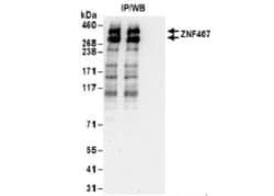
Immunogen
The immunogen this antibody was made to, maps to a region between residue 2000 to 2050 of human Zinc Finger Protein 407 using the numbering given in entry NP_060227.2 (GeneID 55628).
Quantity
100 μL
Primary or Secondary
Primary
Test Specificity
Based on 100% sequence identity, this antibody is predicted to react with Orangutan, Gorilla and Chimpanzee.
Content And Storage
Store at 4C short term. Aliquot and store at -20C long term. Avoid freeze-thaw cycles.
Related Products

Description
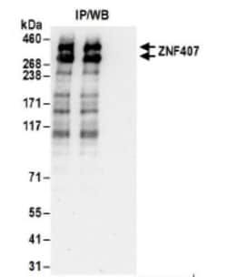
- Description ZNF407 Polyclonal specifically detects ZNF407 in Human samples
- It is validated for Immunoprecipitation.
Compare Similar Items
Show Difference
Antigen: ZNF407
Classification: Polyclonal
Conjugate: Unconjugated
Formulation: Tris-Citrate/Phosphate (pH 7.0 - 8.0) with 0.09% Sodium Azide
Gene Alias: FLJ13839, FLJ20307, KIAA1703, zinc finger protein 407
Host Species: Rabbit
Purification Method: Affinity Purified
Regulatory Status: RUO
Gene ID (Entrez): 55628
Target Species: Human
Isotype: IgG
Applications: Immunoprecipitation
Concentration: 1.0 mg/mL
Dilution: Immunoprecipitation 2 - 10 ug/mg lysate
Gene Accession No.: NP_060227.2
Gene Symbols: ZNF407
Immunogen: The immunogen this antibody was made to, maps to a region between residue 2000 to 2050 of human Zinc Finger Protein 407 using the numbering given in entry NP_060227.2 (GeneID 55628).
Quantity: 100 μL
Primary or Secondary: Primary
Test Specificity: Based on 100% sequence identity, this antibody is predicted to react with Orangutan, Gorilla and Chimpanzee.
Content And Storage: Store at 4C short term. Aliquot and store at -20C long term. Avoid freeze-thaw cycles.

Antigen: CEACAM5/CD66e
Classification: Monoclonal
Conjugate: DyLight 650
Formulation: __
Gene Alias: Carcinoembryonic antigen, carcinoembryonic antigen-related cell adhesion molecule 5, CD66e antigen, CEACD66e, DKFZp781M2392, Meconium antigen 100
Host Species: Mouse
Purification Method: Protein A or G purified
Regulatory Status: RUO
Gene ID (Entrez): 1048
Target Species: Human
Isotype: IgG2a κ
Applications: Western Blot, Flow Cytometry, ELISA, Immunocytochemistry, Immunofluorescence, Immunohistochemistry (Paraffin)
Concentration: __
Dilution: Western Blot, Flow Cytometry, ELISA, Immunocytochemistry/Immunofluorescence, Immunohistochemistry-Paraffin, Immunohistochemistry-Frozen
Gene Accession No.: __
Gene Symbols: CEACAM5
Immunogen: Human colon carcinoma extract (Uniprot: P06731)
Quantity: 0.1 mL
Primary or Secondary: Primary
Test Specificity: This antibody recognizes proteins of 80-200kDa, identified as different members of CEA family. CEA is synthesized during development in the fetal gut and is re-expressed in increased amounts in intestinal carcinomas and several other tumors. This monoclonal antibody does not react with nonspecific cross-reacting antigen (NCA) and with human polymorphonuclear leucocytes. It shows no reaction with a variety of normal tissues and is suitable for staining of formalin/paraffin tissues. CEA is not found in benign glands, stroma, or malignant prostatic cells. Antibody to CEA is useful in detecting early foci of gastric carcinoma and in distinguishing pulmonary adenocarcinomas (60-70% are CEA+) from pleural mesotheliomas (rarely or weakly CEA+). Anti-CEA positivity is seen in adenocarcinomas from the lung, colon, stomach, esophagus, pancreas, gallbladder, urachus, salivary gland, ovary, and endocervix.
Content And Storage: Store at 4C in the dark.
Antigen: CEACAM5/CD66e
Classification: Monoclonal
Conjugate: DyLight 680
Formulation: __
Gene Alias: Carcinoembryonic antigen, carcinoembryonic antigen-related cell adhesion molecule 5, CD66e antigen, CEACD66e, DKFZp781M2392, Meconium antigen 100
Host Species: Mouse
Purification Method: Protein A or G purified
Regulatory Status: RUO
Gene ID (Entrez): 1048
Target Species: Human
Isotype: IgG2a κ
Applications: Western Blot, Flow Cytometry, ELISA, Immunocytochemistry, Immunofluorescence, Immunohistochemistry (Paraffin)
Concentration: __
Dilution: Western Blot, Flow Cytometry, ELISA, Immunocytochemistry/Immunofluorescence, Immunohistochemistry-Paraffin, Immunohistochemistry-Frozen
Gene Accession No.: __
Gene Symbols: CEACAM5
Immunogen: Human colon carcinoma extract (Uniprot: P06731)
Quantity: 0.1 mL
Primary or Secondary: Primary
Test Specificity: This antibody recognizes proteins of 80-200kDa, identified as different members of CEA family. CEA is synthesized during development in the fetal gut and is re-expressed in increased amounts in intestinal carcinomas and several other tumors. This monoclonal antibody does not react with nonspecific cross-reacting antigen (NCA) and with human polymorphonuclear leucocytes. It shows no reaction with a variety of normal tissues and is suitable for staining of formalin/paraffin tissues. CEA is not found in benign glands, stroma, or malignant prostatic cells. Antibody to CEA is useful in detecting early foci of gastric carcinoma and in distinguishing pulmonary adenocarcinomas (60-70% are CEA+) from pleural mesotheliomas (rarely or weakly CEA+). Anti-CEA positivity is seen in adenocarcinomas from the lung, colon, stomach, esophagus, pancreas, gallbladder, urachus, salivary gland, ovary, and endocervix.
Content And Storage: Store at 4C in the dark.