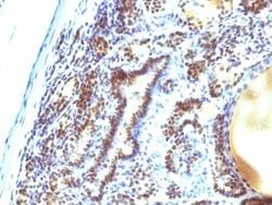
TRIM29 Antibody (TRIM29/1042), Novus Biologicals™
Manufacturer: Fischer Scientific
The price for this product is unavailable. Please request a quote
Antigen
TRIM29
Classification
Monoclonal
Concentration
0.2mg/mL
Dilution
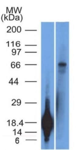
Western Blot 0.5 - 1.0 ug/ml, Flow Cytometry 0.5 - 1 ug/million cells in 0.1 ml, SDS-Page, Immunofluorescence 0.5 - 1.0 ug/ml
Gene Accession No.
Q14134
Gene Symbols
TRIM29
Immunogen
Recombinant fragment (126 Amino acid residues between aa 1-200) of human TRIM29 protein
Purification Method
Protein A or G purified
Regulatory Status
RUO
Gene ID (Entrez)
23650
Target Species
Human
Form
Purified
Applications
Western Blot, Flow Cytometry, SDS-Page, Immunofluorescence
Clone
TRIM29/1042
Conjugate
Unconjugated
Formulation
1.0mM PBS and 0.05% BSA with 0.05% Sodium Azide
Gene Alias
Ataxia telangiectasia group D-associated protein, ATDCataxia-telangiectasia group D-associated protein, FLJ36085, tripartite motif containing 29, tripartite motif protein TRIM29, tripartite motif-containing 29, tripartite motif-containing protein 29
Host Species
Mouse
Molecular Weight of Antigen
66 kDa
Quantity
0.2 mg
Primary or Secondary
Primary
Test Specificity
It recognizes a 66kDa protein, which is identified as Tripartite motif-containing protein 29 (TRIM29). It interacts with the intermediate filament protein vimentin, a substrate for the PKC family of protein kinases, and with hPKCI-1, an inhibitor of the PKCs. TRIM29 protein contains both zinc finger and leucine zipper motifs, suggesting that the it may form homodimers and possibly associate with DNA. High expression of TRIM29 has been reported in gastric cancer and pancreatic cancer, and correlates with enhanced tumor growth and lymph node metastasis. TRIM29 is also able to distinguish lung squamous cell carcinoma from lung adenocarcinoma with ∼90% positive accuracy, when used in a panel with TTF-1, p63, CK5/6, and Napsin-A antibodies.
Content And Storage
Store at 4C.
Isotype
IgG2b κ
Description
- Ensure accurate, reproducible results in Western Blot, Flow Cytometry, Immunofluorescence TRIM29 Monoclonal specifically detects TRIM29 in Human samples
- It is validated for Western Blot.
Compare Similar Items
Show Difference
Antigen: TRIM29
Classification: Monoclonal
Concentration: 0.2mg/mL
Dilution: Western Blot 0.5 - 1.0 ug/ml, Flow Cytometry 0.5 - 1 ug/million cells in 0.1 ml, SDS-Page, Immunofluorescence 0.5 - 1.0 ug/ml
Gene Accession No.: Q14134
Gene Symbols: TRIM29
Immunogen: Recombinant fragment (126 Amino acid residues between aa 1-200) of human TRIM29 protein
Purification Method: Protein A or G purified
Regulatory Status: RUO
Gene ID (Entrez): 23650
Target Species: Human
Form: Purified
Applications: Western Blot, Flow Cytometry, SDS-Page, Immunofluorescence
Clone: TRIM29/1042
Conjugate: Unconjugated
Formulation: 1.0mM PBS and 0.05% BSA with 0.05% Sodium Azide
Gene Alias: Ataxia telangiectasia group D-associated protein, ATDCataxia-telangiectasia group D-associated protein, FLJ36085, tripartite motif containing 29, tripartite motif protein TRIM29, tripartite motif-containing 29, tripartite motif-containing protein 29
Host Species: Mouse
Molecular Weight of Antigen: 66 kDa
Quantity: 0.2 mg
Primary or Secondary: Primary
Test Specificity: It recognizes a 66kDa protein, which is identified as Tripartite motif-containing protein 29 (TRIM29). It interacts with the intermediate filament protein vimentin, a substrate for the PKC family of protein kinases, and with hPKCI-1, an inhibitor of the PKCs. TRIM29 protein contains both zinc finger and leucine zipper motifs, suggesting that the it may form homodimers and possibly associate with DNA. High expression of TRIM29 has been reported in gastric cancer and pancreatic cancer, and correlates with enhanced tumor growth and lymph node metastasis. TRIM29 is also able to distinguish lung squamous cell carcinoma from lung adenocarcinoma with ∼90% positive accuracy, when used in a panel with TTF-1, p63, CK5/6, and Napsin-A antibodies.
Content And Storage: Store at 4C.
Isotype: IgG2b κ

Antigen: MEKK1
Classification: Monoclonal
Concentration: 0.2mg/mL
Dilution: Flow Cytometry 0.5 - 1 ug/million cells in 0.1 ml, Immunohistochemistry-Paraffin 0.5 - 1.0 ug/ml, Immunofluorescence 1 - 2 ug/ml
Gene Accession No.: Q13233
Gene Symbols: MAP3K1
Immunogen: Partial recombinant MAP3K1 (aa1211-1310) (SKNSMTLDLNSSSKCDDSFGCSSNSSNAVIPSDETVFTP-VEEKCRLDVNTELNSSIEDLLEASMPSSDTTVTFKSEVAVLSPEKAENDDTYKDDVNHNQK)
Purification Method: Protein A or G purified
Regulatory Status: RUO
Gene ID (Entrez): 4214
Target Species: Human
Form: Purified
Applications: Flow Cytometry, Immunohistochemistry (Paraffin), Immunofluorescence
Clone: 2F6
Conjugate: Unconjugated
Formulation: 10mM PBS and 0.05% BSA with 0.05% Sodium Azide
Gene Alias: MAPK/ERK kinase kinase 1, MAPKKK1MAP/ERK kinase kinase 1, MEK kinase 1, MEKK1EC 2.7.11.25, MEKKMEKK 1, mitogen-activated protein kinase kinase kinase 1
Host Species: Mouse
Molecular Weight of Antigen: __
Quantity: 0.02 mg
Primary or Secondary: Primary
Test Specificity: Mitogen-activated protein (MAP) kinase cascades are activated by various extracellular stimuli, including growth factors. The MEK kinases (also designated MAP kinase kinase kinases, MKKKs, MAP3Ks or MEKKs) phosphorylate and thereby activate the MEKs (also called MAP kinase kinases or MKKs), including ERK, JNK and p38. These activated MEKs in turn phosphorylate and activate the MAP kinases. The MEK kinases include Raf-1, Raf-B, Mos, MEK kinase-1, MEK kinase-2, MEK kinase-3, MEK kinase-4 and ASK 1 (MEK kinase- 5). MEK kinase-1 activates the ERK and c-Jun NH2-terminal kinase (JNK) pathways by phosphorylation of MAP2K1 and MAP2K4, and also activates the central protein kinases of the NF B pathway, CHUK and IKBKB. Additionally, MEK kinase-1 uses an E3 ligase through its PHD domain, a RING-finger-like structure, to target proteins for degradation through ubiquitination.
Content And Storage: Store at 4C.
Isotype: IgG2a κ
Antigen: MEKK1
Classification: Monoclonal
Concentration: 0.2mg/mL
Dilution: Flow Cytometry 0.5 - 1 ug/million cells in 0.1 ml, Immunohistochemistry-Paraffin 0.5 - 1.0 ug/ml, Immunofluorescence 1 - 2 ug/ml
Gene Accession No.: Q13233
Gene Symbols: MAP3K1
Immunogen: Partial recombinant MAP3K1 (aa1211-1310) (SKNSMTLDLNSSSKCDDSFGCSSNSSNAVIPSDETVFTP-VEEKCRLDVNTELNSSIEDLLEASMPSSDTTVTFKSEVAVLSPEKAENDDTYKDDVNHNQK)
Purification Method: Protein A or G purified
Regulatory Status: RUO
Gene ID (Entrez): 4214
Target Species: Human
Form: Purified
Applications: Flow Cytometry, Immunohistochemistry (Paraffin), Immunofluorescence
Clone: 2F6
Conjugate: Unconjugated
Formulation: 10mM PBS and 0.05% BSA with 0.05% Sodium Azide
Gene Alias: MAPK/ERK kinase kinase 1, MAPKKK1MAP/ERK kinase kinase 1, MEK kinase 1, MEKK1EC 2.7.11.25, MEKKMEKK 1, mitogen-activated protein kinase kinase kinase 1
Host Species: Mouse
Molecular Weight of Antigen: __
Quantity: 0.1 mg
Primary or Secondary: Primary
Test Specificity: Mitogen-activated protein (MAP) kinase cascades are activated by various extracellular stimuli, including growth factors. The MEK kinases (also designated MAP kinase kinase kinases, MKKKs, MAP3Ks or MEKKs) phosphorylate and thereby activate the MEKs (also called MAP kinase kinases or MKKs), including ERK, JNK and p38. These activated MEKs in turn phosphorylate and activate the MAP kinases. The MEK kinases include Raf-1, Raf-B, Mos, MEK kinase-1, MEK kinase-2, MEK kinase-3, MEK kinase-4 and ASK 1 (MEK kinase- 5). MEK kinase-1 activates the ERK and c-Jun NH2-terminal kinase (JNK) pathways by phosphorylation of MAP2K1 and MAP2K4, and also activates the central protein kinases of the NF B pathway, CHUK and IKBKB. Additionally, MEK kinase-1 uses an E3 ligase through its PHD domain, a RING-finger-like structure, to target proteins for degradation through ubiquitination.
Content And Storage: Store at 4C.
Isotype: IgG2a κ