Caldesmon/CALD1 Antibody (CALD1/820), Novus Biologicals™
Manufacturer: Fischer Scientific
Select a Size
| Pack Size | SKU | Availability | Price |
|---|---|---|---|
| Each of 1 | NBP24442300-Each-of-1 | In Stock | ₹ 23,585.00 |
NBP24442300 - Each of 1
In Stock
Quantity
1
Base Price: ₹ 23,585.00
GST (18%): ₹ 4,245.30
Total Price: ₹ 27,830.30
Antigen
Caldesmon/CALD1
Classification
Monoclonal
Concentration
0.2mg/mL
Dilution
Flow Cytometry 0.5 - 1 ug/million cells in 0.1 ml, Immunohistochemistry-Paraffin 0.25 - 0.5 ug/ml, Immunofluorescence 1 - 2 ug/ml
Gene Accession No.
Q05682
Gene Symbols
CALD1
Immunogen
Recombinant full-length human CALD1 protein
Purification Method
Protein A or G purified
Regulatory Status
RUO
Primary or Secondary
Primary
Test Specificity
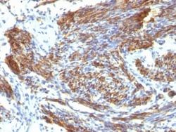
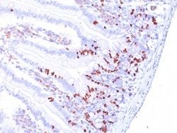
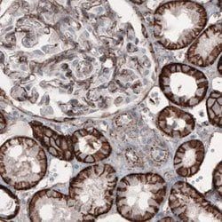
Recognizes a protein of 150kDa, which is identified as the high molecular weight variant of Caldesmon. Two closely related variants of human caldesmon have been identified which are different in their electrophoretic mobility and cellular distribution. The h-caldesmon variant (120-150kDa) is predominantly expressed in smooth muscle whereas l-caldesmon (70-80kDa) is found in non- muscle tissue and cells. Neither of the two variants has been detected in skeletal muscle. This MAb recognizes only the 150kDa variant (h-caldesmon) in Western blots of human aortic media extracts and is unreactive with fibroblast extracts from cultivated human foreskin. Caldesmon is a developmentally regulated protein involved in smooth muscle and non-muscle contraction.
Content And Storage
Store at 4C.
Isotype
IgG1 κ
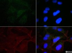
Applications
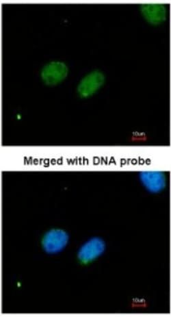
Flow Cytometry, Immunohistochemistry (Paraffin), Immunofluorescence
Clone
CALD1/820
Conjugate
Unconjugated
Formulation
10mM PBS and 0.05% BSA with 0.05% Sodium Azide
Gene Alias
CAD, caldesmon, caldesmon 1, CDMH-CAD, HCAD, LCAD, L-CAD, MGC21352, NAG22
Host Species
Mouse
Molecular Weight of Antigen
150 kDa
Quantity
0.02 mg
Research Discipline
Cytoskeleton Markers
Gene ID (Entrez)
800
Target Species
Human, Rat (Negative)
Form
Purified
Related Products


Description
- Ensure accurate, reproducible results in Flow Cytometry, Immunohistochemistry (Paraffin), Immunofluorescence Caldesmon/CALD1 Monoclonal specifically detects Caldesmon/CALD1 in Human, Rat (Negative) samples
- It is validated for Western Blot, Immunohistochemistry, Immunohistochemistry-Paraffin.
Compare Similar Items
Show Difference
Antigen: Caldesmon/CALD1
Classification: Monoclonal
Concentration: 0.2mg/mL
Dilution: Flow Cytometry 0.5 - 1 ug/million cells in 0.1 ml, Immunohistochemistry-Paraffin 0.25 - 0.5 ug/ml, Immunofluorescence 1 - 2 ug/ml
Gene Accession No.: Q05682
Gene Symbols: CALD1
Immunogen: Recombinant full-length human CALD1 protein
Purification Method: Protein A or G purified
Regulatory Status: RUO
Primary or Secondary: Primary
Test Specificity: Recognizes a protein of 150kDa, which is identified as the high molecular weight variant of Caldesmon. Two closely related variants of human caldesmon have been identified which are different in their electrophoretic mobility and cellular distribution. The h-caldesmon variant (120-150kDa) is predominantly expressed in smooth muscle whereas l-caldesmon (70-80kDa) is found in non- muscle tissue and cells. Neither of the two variants has been detected in skeletal muscle. This MAb recognizes only the 150kDa variant (h-caldesmon) in Western blots of human aortic media extracts and is unreactive with fibroblast extracts from cultivated human foreskin. Caldesmon is a developmentally regulated protein involved in smooth muscle and non-muscle contraction.
Content And Storage: Store at 4C.
Isotype: IgG1 κ
Applications: Flow Cytometry, Immunohistochemistry (Paraffin), Immunofluorescence
Clone: CALD1/820
Conjugate: Unconjugated
Formulation: 10mM PBS and 0.05% BSA with 0.05% Sodium Azide
Gene Alias: CAD, caldesmon, caldesmon 1, CDMH-CAD, HCAD, LCAD, L-CAD, MGC21352, NAG22
Host Species: Mouse
Molecular Weight of Antigen: 150 kDa
Quantity: 0.02 mg
Research Discipline: Cytoskeleton Markers
Gene ID (Entrez): 800
Target Species: Human, Rat (Negative)
Form: Purified
Antigen: Caldesmon/CALD1
Classification: Monoclonal
Concentration: 0.2mg/mL
Dilution: Flow Cytometry 0.5 - 1 ug/million cells in 0.1 ml, Immunohistochemistry-Paraffin 0.25 - 0.5 ug/ml, Immunofluorescence 1 - 2 ug/ml
Gene Accession No.: Q05682
Gene Symbols: CALD1
Immunogen: Recombinant full-length human CALD1 protein
Purification Method: Protein A or G purified
Regulatory Status: RUO
Primary or Secondary: Primary
Test Specificity: Recognizes a protein of 150kDa, which is identified as the high molecular weight variant of Caldesmon. Two closely related variants of human caldesmon have been identified which are different in their electrophoretic mobility and cellular distribution. The h-caldesmon variant (120-150kDa) is predominantly expressed in smooth muscle whereas l-caldesmon (70-80kDa) is found in non- muscle tissue and cells. Neither of the two variants has been detected in skeletal muscle. This MAb recognizes only the 150kDa variant (h-caldesmon) in Western blots of human aortic media extracts and is unreactive with fibroblast extracts from cultivated human foreskin. Caldesmon is a developmentally regulated protein involved in smooth muscle and non-muscle contraction.
Content And Storage: Store at 4C.
Isotype: IgG1 κ
Applications: Flow Cytometry, Immunohistochemistry (Paraffin), Immunofluorescence
Clone: CALD1/820
Conjugate: Unconjugated
Formulation: 10mM PBS and 0.05% BSA with 0.05% Sodium Azide
Gene Alias: CAD, caldesmon, caldesmon 1, CDMH-CAD, HCAD, LCAD, L-CAD, MGC21352, NAG22
Host Species: Mouse
Molecular Weight of Antigen: 150 kDa
Quantity: 0.1 mg
Research Discipline: Cytoskeleton Markers
Gene ID (Entrez): 800
Target Species: Human, Rat (Negative)
Form: Purified
Antigen: Caldesmon/CALD1
Classification: Monoclonal
Concentration: 0.2mg/mL
Dilution: Flow Cytometry 0.5 - 1 ug/million cells in 0.1 ml, Immunohistochemistry-Paraffin 0.25 - 0.5 ug/ml, Immunofluorescence 1 - 2 ug/ml
Gene Accession No.: Q05682
Gene Symbols: CALD1
Immunogen: Recombinant full-length human CALD1 protein
Purification Method: Protein A or G purified
Regulatory Status: RUO
Primary or Secondary: Primary
Test Specificity: Recognizes a protein of 150kDa, which is identified as the high molecular weight variant of Caldesmon. Two closely related variants of human caldesmon have been identified which are different in their electrophoretic mobility and cellular distribution. The h-caldesmon variant (120-150kDa) is predominantly expressed in smooth muscle whereas l-caldesmon (70-80kDa) is found in non- muscle tissue and cells. Neither of the two variants has been detected in skeletal muscle. This MAb recognizes only the 150kDa variant (h-caldesmon) in Western blots of human aortic media extracts and is unreactive with fibroblast extracts from cultivated human foreskin. Caldesmon is a developmentally regulated protein involved in smooth muscle and non-muscle contraction.
Content And Storage: Store at 4C.
Isotype: IgG1 κ
Applications: Flow Cytometry, Immunohistochemistry (Paraffin), Immunofluorescence
Clone: CALD1/820
Conjugate: Unconjugated
Formulation: 10mM PBS and 0.05% BSA with 0.05% Sodium Azide
Gene Alias: CAD, caldesmon, caldesmon 1, CDMH-CAD, HCAD, LCAD, L-CAD, MGC21352, NAG22
Host Species: Mouse
Molecular Weight of Antigen: 150 kDa
Quantity: 0.2 mg
Research Discipline: Cytoskeleton Markers
Gene ID (Entrez): 800
Target Species: Human, Rat (Negative)
Form: Purified