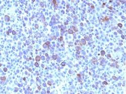
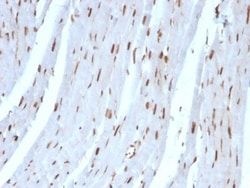
SUMO2 Antibody (SUMO2/1199), Novus Biologicals™
Manufacturer: Fischer Scientific
The price for this product is unavailable. Please request a quote
Antigen
SUMO2
Classification
Monoclonal
Concentration
0.2mg/mL
Dilution
Flow Cytometry 0.5 - 1 ug/million cells in 0.1 ml, Immunohistochemistry-Paraffin 0.5 - 1.0 ug/ml, Immunofluorescence 0.5 - 1.0 ug/ml
Gene Accession No.
P55854
Gene Symbols
SUMO2
Immunogen
Recombinant human SUMO2 protein
Purification Method
Protein A or G purified
Regulatory Status
RUO
Gene ID (Entrez)
6613
Target Species
Human, Rat
Form
Purified
Applications
Flow Cytometry, Immunohistochemistry (Paraffin), Immunofluorescence
Clone
SUMO2/1199
Conjugate
Unconjugated
Formulation
10mM PBS and 0.05% BSA with 0.05% Sodium Azide
Gene Alias
HSMT3, MGC117191, sentrin 2, Sentrin-2, small ubiquitin-like modifier 2, small ubiquitin-related modifier 2, SMT3 (suppressor of mif two 3, yeast) homolog 2, SMT3 homolog 2, SMT3 suppressor of mif two 3 homolog 2 (S. cerevisiae), SMT3 suppressor of mif two 3 homolog 2 (yeast), SMT3A, SMT3B, SMT3H2, SUMO-2, SUMO3, SUMO-3, Ubiquitin-like protein SMT3A, ubiquitin-like protein SMT3B
Host Species
Mouse
Molecular Weight of Antigen
12 kDa
Quantity
0.2 mg
Primary or Secondary
Primary
Test Specificity
The small ubiquitin-related modifier (SUMO) proteins, which include SUMO-1, 2 and 3, belong to the ubiquitin-like protein family. Like ubiquitin, the SUMO proteins are synthesized as precursor proteins that undergo processing before conjugation to target proteins. Also, both utilize the E1, E2 and E3 cascade enzymes for conjugation. However, SUMO and ubiquitin differ with respect to targeting. Ubiquitination predominantly targets proteins for degradation, whereas sumoylation targets proteins to a variety of cellular processing, including nuclear transport, transcriptional regulation, apoptosis and protein stability. The unconjugated SUMO-1, 2 and 3 proteins localize to the nuclear membrane, nuclear bodies and cytoplasm, respectively. SUMO-1 utilizes Ubc9 for conjugation to several target proteins, which include MDM2, p53, PML and RanGap1. SUMO-2 and 3 contribute to a greater percentage of protein modification than does SUMO-1 and unlike SUMO-1, they can form polymeric chains. In additi
Content And Storage
Store at 4C.
Isotype
IgG1 κ
Description
- Ensure accurate, reproducible results in Flow Cytometry, Immunohistochemistry (Paraffin), Immunofluorescence SUMO2 Monoclonal specifically detects SUMO2 in Human, Rat samples
- It is validated for Flow Cytometry, Immunohistochemistry, Immunocytochemistry/Immunofluorescence, Immunohistochemistry-Paraffin, Immunofluorescence.
Compare Similar Items
Show Difference
Antigen: SUMO2
Classification: Monoclonal
Concentration: 0.2mg/mL
Dilution: Flow Cytometry 0.5 - 1 ug/million cells in 0.1 ml, Immunohistochemistry-Paraffin 0.5 - 1.0 ug/ml, Immunofluorescence 0.5 - 1.0 ug/ml
Gene Accession No.: P55854
Gene Symbols: SUMO2
Immunogen: Recombinant human SUMO2 protein
Purification Method: Protein A or G purified
Regulatory Status: RUO
Gene ID (Entrez): 6613
Target Species: Human, Rat
Form: Purified
Applications: Flow Cytometry, Immunohistochemistry (Paraffin), Immunofluorescence
Clone: SUMO2/1199
Conjugate: Unconjugated
Formulation: 10mM PBS and 0.05% BSA with 0.05% Sodium Azide
Gene Alias: HSMT3, MGC117191, sentrin 2, Sentrin-2, small ubiquitin-like modifier 2, small ubiquitin-related modifier 2, SMT3 (suppressor of mif two 3, yeast) homolog 2, SMT3 homolog 2, SMT3 suppressor of mif two 3 homolog 2 (S. cerevisiae), SMT3 suppressor of mif two 3 homolog 2 (yeast), SMT3A, SMT3B, SMT3H2, SUMO-2, SUMO3, SUMO-3, Ubiquitin-like protein SMT3A, ubiquitin-like protein SMT3B
Host Species: Mouse
Molecular Weight of Antigen: 12 kDa
Quantity: 0.2 mg
Primary or Secondary: Primary
Test Specificity: The small ubiquitin-related modifier (SUMO) proteins, which include SUMO-1, 2 and 3, belong to the ubiquitin-like protein family. Like ubiquitin, the SUMO proteins are synthesized as precursor proteins that undergo processing before conjugation to target proteins. Also, both utilize the E1, E2 and E3 cascade enzymes for conjugation. However, SUMO and ubiquitin differ with respect to targeting. Ubiquitination predominantly targets proteins for degradation, whereas sumoylation targets proteins to a variety of cellular processing, including nuclear transport, transcriptional regulation, apoptosis and protein stability. The unconjugated SUMO-1, 2 and 3 proteins localize to the nuclear membrane, nuclear bodies and cytoplasm, respectively. SUMO-1 utilizes Ubc9 for conjugation to several target proteins, which include MDM2, p53, PML and RanGap1. SUMO-2 and 3 contribute to a greater percentage of protein modification than does SUMO-1 and unlike SUMO-1, they can form polymeric chains. In additi
Content And Storage: Store at 4C.
Isotype: IgG1 κ

Antigen: Glypican 3
Classification: Monoclonal
Concentration: 0.2mg/mL
Dilution: Flow Cytometry 0.5 - 1 ug/million cells in 0.1 ml, Immunohistochemistry-Paraffin 0.5 - 1.0 ug/ml, Immunofluorescence 0.5 - 1.0 ug/ml
Gene Accession No.: P51654
Gene Symbols: GPC3
Immunogen: A recombinant fragment containing amino acids 511-580 of human glypican-3.
Purification Method: Protein A or G purified
Regulatory Status: RUO
Gene ID (Entrez): 2719
Target Species: Human
Form: Purified
Applications: Flow Cytometry, Immunohistochemistry (Paraffin), Immunofluorescence
Clone: SPM595
Conjugate: Unconjugated
Formulation: 1.0mM PBS and 0.05% BSA with 0.05% Sodium Azide
Gene Alias: DGSX, glypican 3, glypican proteoglycan 3, glypican-3, GTR2-2, heparan sulphate proteoglycan, Intestinal protein OCI-5, MXR7, OCI5, OCI-5, secreted glypican-3, SGB, SGBS, SGBS1SDYS
Host Species: Mouse
Molecular Weight of Antigen: 67 kDa
Quantity: 0.02 mg
Primary or Secondary: Primary
Test Specificity: Glypican-3 (GPC3) is a glycosylphospatidyl inositol-anchored membrane protein, which may also be found in a secreted form. Anti-GPC3 has been identified as a useful tumor marker for the diagnosis of hepatocellular carcinoma (HCC), hepatoblastoma, melanoma, testicular germ cell tumors, and Wilm s tumor. In patients with HCC, GPC3 is overexpressed in neoplastic liver tissue and elevated in serum, but is undetectable in normal liver, benign liver, and the serum of healthy donors. GPC3 expression is also found to be higher in HCC liver tissue than in cirrhotic liver or liver with focal lesions such as dysplastic nodules and areas of hepatic adenoma (HA) with malignant transformation. In the context of testicular germ cell tumors, GPC3 expression is up regulated in certain histologic subtypes, specifically yolk sac tumors and choriocarcinoma. A high level of GPC3 expression is also found in some types of embryonal tumors, such as Wilm s tumor and hepatoblastoma, with a low or undetectable e
Content And Storage: Store at 4C.
Isotype: IgG1 κ
Antigen: Glypican 3
Classification: Monoclonal
Concentration: 0.2mg/mL
Dilution: Flow Cytometry 0.5 - 1 ug/million cells in 0.1 ml, Immunohistochemistry-Paraffin 0.5 - 1.0 ug/ml, Immunofluorescence 0.5 - 1.0 ug/ml
Gene Accession No.: P51654
Gene Symbols: GPC3
Immunogen: A recombinant fragment containing amino acids 511-580 of human glypican-3.
Purification Method: Protein A or G purified
Regulatory Status: RUO
Gene ID (Entrez): 2719
Target Species: Human
Form: Purified
Applications: Flow Cytometry, Immunohistochemistry (Paraffin), Immunofluorescence
Clone: SPM595
Conjugate: Unconjugated
Formulation: 1.0mM PBS and 0.05% BSA with 0.05% Sodium Azide
Gene Alias: DGSX, glypican 3, glypican proteoglycan 3, glypican-3, GTR2-2, heparan sulphate proteoglycan, Intestinal protein OCI-5, MXR7, OCI5, OCI-5, secreted glypican-3, SGB, SGBS, SGBS1SDYS
Host Species: Mouse
Molecular Weight of Antigen: 67 kDa
Quantity: 0.1 mg
Primary or Secondary: Primary
Test Specificity: Glypican-3 (GPC3) is a glycosylphospatidyl inositol-anchored membrane protein, which may also be found in a secreted form. Anti-GPC3 has been identified as a useful tumor marker for the diagnosis of hepatocellular carcinoma (HCC), hepatoblastoma, melanoma, testicular germ cell tumors, and Wilm s tumor. In patients with HCC, GPC3 is overexpressed in neoplastic liver tissue and elevated in serum, but is undetectable in normal liver, benign liver, and the serum of healthy donors. GPC3 expression is also found to be higher in HCC liver tissue than in cirrhotic liver or liver with focal lesions such as dysplastic nodules and areas of hepatic adenoma (HA) with malignant transformation. In the context of testicular germ cell tumors, GPC3 expression is up regulated in certain histologic subtypes, specifically yolk sac tumors and choriocarcinoma. A high level of GPC3 expression is also found in some types of embryonal tumors, such as Wilm s tumor and hepatoblastoma, with a low or undetectable e
Content And Storage: Store at 4C.
Isotype: IgG1 κ