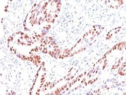
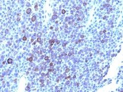
Glypican 3 Antibody (1G12 + GPC3/863), Novus Biologicals™
Manufacturer: Fischer Scientific
The price for this product is unavailable. Please request a quote
Antigen
Glypican 3
Classification
Monoclonal
Concentration
0.2mg/mL
Dilution
Flow Cytometry 0.5 - 1 ug/million cells in 0.1 ml, Immunohistochemistry-Paraffin 0.5 - 1.0 ug/ml, Immunofluorescence 0.5 - 1.0 ug/ml
Gene Accession No.
P51654, P51654
Gene Symbols
GPC3
Immunogen
Recombinant fragment containing amino acids 511-580 of human glypican-3 (1G12); Recombinant full-length human GPC3 protein (GPC3/863)
Purification Method
Protein A or G purified
Regulatory Status
RUO
Gene ID (Entrez)
2719
Target Species
Human
Form
Purified
Applications
Flow Cytometry, Immunohistochemistry (Paraffin), Immunofluorescence
Clone
1G12 + GPC3/863
Conjugate
Unconjugated
Formulation
1.0mM PBS and 0.05% BSA with 0.05% Sodium Azide
Gene Alias
DGSX, glypican 3, glypican proteoglycan 3, glypican-3, GTR2-2, heparan sulphate proteoglycan, Intestinal protein OCI-5, MXR7, OCI5, OCI-5, secreted glypican-3, SGB, SGBS, SGBS1SDYS
Host Species
Mouse
Molecular Weight of Antigen
67 kDa
Quantity
0.2 mg
Primary or Secondary
Primary
Test Specificity
Glypican-3 (GPC3) is an integral membrane protein that is mutated in the Simpson-Golabi-Behmel syndrome (SGBS). SGBS is characterized by pre- and post-natal overgrowth and is a recessive X-linked condition. GPC3 may also be found in a secreted form. Anti-GPC3 has been identified as a useful tumor marker for the diagnosis of hepatocellular carcinoma (HCC), hepatoblastoma, melanoma, testicular germ cell tumors, and Wilm s tumor. In patients with HCC, GPC3 is overexpressed in neoplastic liver tissue and elevated in serum, but is undetectable in normal liver, benign liver, and the serum of healthy donors. GPC3 expression is also found to be higher in HCC liver tissue than in cirrhotic liver or liver with focal lesions such as dysplastic nodules and areas of hepatic adenoma (HA) with malignant transformation. In the context of testicular germ cell tumors, GPC3 expression is up regulated in certain histologic subtypes, specifically yolk sac tumors and choriocarcinoma. A high level of GPC3 ex
Content And Storage
Store at 4C.
Isotype
IgG
Description
- Ensure accurate, reproducible results in Flow Cytometry, Immunohistochemistry (Paraffin), Immunofluorescence Glypican 3 Monoclonal specifically detects Glypican 3 in Human samples
- It is validated for Flow Cytometry, Immunohistochemistry, Immunocytochemistry/Immunofluorescence, Immunohistochemistry-Paraffin, Immunofluorescence.
Compare Similar Items
Show Difference
Antigen: Glypican 3
Classification: Monoclonal
Concentration: 0.2mg/mL
Dilution: Flow Cytometry 0.5 - 1 ug/million cells in 0.1 ml, Immunohistochemistry-Paraffin 0.5 - 1.0 ug/ml, Immunofluorescence 0.5 - 1.0 ug/ml
Gene Accession No.: P51654, P51654
Gene Symbols: GPC3
Immunogen: Recombinant fragment containing amino acids 511-580 of human glypican-3 (1G12); Recombinant full-length human GPC3 protein (GPC3/863)
Purification Method: Protein A or G purified
Regulatory Status: RUO
Gene ID (Entrez): 2719
Target Species: Human
Form: Purified
Applications: Flow Cytometry, Immunohistochemistry (Paraffin), Immunofluorescence
Clone: 1G12 + GPC3/863
Conjugate: Unconjugated
Formulation: 1.0mM PBS and 0.05% BSA with 0.05% Sodium Azide
Gene Alias: DGSX, glypican 3, glypican proteoglycan 3, glypican-3, GTR2-2, heparan sulphate proteoglycan, Intestinal protein OCI-5, MXR7, OCI5, OCI-5, secreted glypican-3, SGB, SGBS, SGBS1SDYS
Host Species: Mouse
Molecular Weight of Antigen: 67 kDa
Quantity: 0.2 mg
Primary or Secondary: Primary
Test Specificity: Glypican-3 (GPC3) is an integral membrane protein that is mutated in the Simpson-Golabi-Behmel syndrome (SGBS). SGBS is characterized by pre- and post-natal overgrowth and is a recessive X-linked condition. GPC3 may also be found in a secreted form. Anti-GPC3 has been identified as a useful tumor marker for the diagnosis of hepatocellular carcinoma (HCC), hepatoblastoma, melanoma, testicular germ cell tumors, and Wilm s tumor. In patients with HCC, GPC3 is overexpressed in neoplastic liver tissue and elevated in serum, but is undetectable in normal liver, benign liver, and the serum of healthy donors. GPC3 expression is also found to be higher in HCC liver tissue than in cirrhotic liver or liver with focal lesions such as dysplastic nodules and areas of hepatic adenoma (HA) with malignant transformation. In the context of testicular germ cell tumors, GPC3 expression is up regulated in certain histologic subtypes, specifically yolk sac tumors and choriocarcinoma. A high level of GPC3 ex
Content And Storage: Store at 4C.
Isotype: IgG

Antigen: p57 Kip2
Classification: Monoclonal
Concentration: 0.2mg/mL
Dilution: Flow Cytometry 0.5 - 1 ug/million cells in 0.1 ml, Immunohistochemistry-Paraffin 0.25 - 0.5 ug/ml, Immunofluorescence 0.5 - 1.0 ug/ml
Gene Accession No.: P49918
Gene Symbols: CDKN1C
Immunogen: Recombinant human p57Kip2 protein
Purification Method: Protein A or G purified
Regulatory Status: RUO
Gene ID (Entrez): 1028
Target Species: Human, Mouse
Form: Purified
Applications: Flow Cytometry, Immunohistochemistry (Paraffin), Immunofluorescence
Clone: KIP2/880
Conjugate: Unconjugated
Formulation: 10mM PBS and 0.05% BSA with 0.05% Sodium Azide
Gene Alias: Beckwith-Wiedemann syndrome, BWS, cyclin-dependent kinase inhibitor 1C, cyclin-dependent kinase inhibitor 1C (p57, Kip2), Cyclin-dependent kinase inhibitor p57, KIP2BWCR, p57, p57Kip2, WBS
Host Species: Mouse
Molecular Weight of Antigen: 57 kDa
Quantity: 0.02 mg
Primary or Secondary: Primary
Test Specificity: Recognizes a protein of 57kDa, identified as p57Kip2. It shows no cross-reaction with p27Kip1. p57Kip2 is a potent tight-binding inhibitor of several G1 cyclin complexes, and is a negative regulator of cell proliferation. Anti-p57 has been used as an aide in identification of complete hydatidiform mole (CHM) (no nuclear labeling of cytotrophoblasts and stromal cells) from partial hydatidiform mole (PHM) in which both cytotrophoblasts and stromal cells stain. The histological differentiation of complete mole, partial mole, and hydropic spontaneous abortion is problematic. Most complete hydatidiform moles are diploid, whereas most partial moles are triploid. Ploidy studies will identify partial moles, but will not differentiate complete moles from non-molar gestations. Complete moles carry a high risk of persistent disease and choriocarcinoma, while partial moles have a very low risk. In normal placenta, many cytotrophoblast nuclei and stromal cells are labeled with this antibody. Simila
Content And Storage: Store at 4C.
Isotype: IgG2b κ
Antigen: p57 Kip2
Classification: Monoclonal
Concentration: 0.2mg/mL
Dilution: Flow Cytometry 0.5 - 1 ug/million cells in 0.1 ml, Immunohistochemistry-Paraffin 0.25 - 0.5 ug/ml, Immunofluorescence 0.5 - 1.0 ug/ml
Gene Accession No.: P49918
Gene Symbols: CDKN1C
Immunogen: Recombinant human p57Kip2 protein
Purification Method: Protein A or G purified
Regulatory Status: RUO
Gene ID (Entrez): 1028
Target Species: Human, Mouse
Form: Purified
Applications: Flow Cytometry, Immunohistochemistry (Paraffin), Immunofluorescence
Clone: KIP2/880
Conjugate: Unconjugated
Formulation: 10mM PBS and 0.05% BSA with 0.05% Sodium Azide
Gene Alias: Beckwith-Wiedemann syndrome, BWS, cyclin-dependent kinase inhibitor 1C, cyclin-dependent kinase inhibitor 1C (p57, Kip2), Cyclin-dependent kinase inhibitor p57, KIP2BWCR, p57, p57Kip2, WBS
Host Species: Mouse
Molecular Weight of Antigen: 57 kDa
Quantity: 0.1 mg
Primary or Secondary: Primary
Test Specificity: Recognizes a protein of 57kDa, identified as p57Kip2. It shows no cross-reaction with p27Kip1. p57Kip2 is a potent tight-binding inhibitor of several G1 cyclin complexes, and is a negative regulator of cell proliferation. Anti-p57 has been used as an aide in identification of complete hydatidiform mole (CHM) (no nuclear labeling of cytotrophoblasts and stromal cells) from partial hydatidiform mole (PHM) in which both cytotrophoblasts and stromal cells stain. The histological differentiation of complete mole, partial mole, and hydropic spontaneous abortion is problematic. Most complete hydatidiform moles are diploid, whereas most partial moles are triploid. Ploidy studies will identify partial moles, but will not differentiate complete moles from non-molar gestations. Complete moles carry a high risk of persistent disease and choriocarcinoma, while partial moles have a very low risk. In normal placenta, many cytotrophoblast nuclei and stromal cells are labeled with this antibody. Simila
Content And Storage: Store at 4C.
Isotype: IgG2b κ