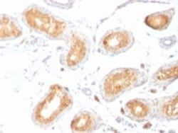
MCAM/CD146 Antibody (SPM620), Novus Biologicals™
Manufacturer: Fischer Scientific
The price for this product is unavailable. Please request a quote
Antigen
MCAM/CD146
Classification
Monoclonal
Concentration
0.2mg/mL
Dilution
Flow Cytometry 0.5 - 1 ug/million cells in 0.1 ml, Immunohistochemistry-Paraffin 0.5 - 1.0 ug/ml, Immunofluorescence 0.5 - 1.0 ug/ml
Gene Alias
CD146, CD146 antigen, cell surface glycoprotein MUC18, Cell surface glycoprotein P1H12, melanoma adhesion molecule, melanoma cell adhesion moleculeS-endo 1 endothelial-associated antigen, Melanoma-associated antigen A32, Melanoma-associated antigen MUC18, MUC18Gicerin
Host Species
Mouse
Molecular Weight of Antigen
130 kDa
Quantity
0.2 mg
Research Discipline
Cancer, Mesenchymal Stem Cell Markers, Stem Cell Markers
Gene ID (Entrez)
4162
Target Species
Human
Form
Purified
Applications
Flow Cytometry, Immunohistochemistry (Paraffin), Immunofluorescence
Clone
SPM620
Conjugate
Unconjugated
Formulation
10mM PBS and 0.05% BSA with 0.05% Sodium Azide
Gene Symbols
MCAM
Immunogen
Recombinant human MCAM protein
Purification Method
Protein A or G purified
Regulatory Status
RUO
Primary or Secondary
Primary
Test Specificity
The human Mel-CAM gene maps to chromosome 11q23 and encodes a trans-membrane glycoprotein, also designated MCAM, MUC 18 or CD146, that belongs to the immunoglobulin superfamily and functions as a Ca2+-independent cell adhesion molecule. Mel-CAM expression is restricted to advanced primary and metastatic melanomas and to cell lines of the neuroectodermal lineage, but not normal melanocytes. Mel-CAM is found on 80% of advanced primary human melanomas and correlates well with development of metastatic disease.
Content And Storage
Store at 4C.
Isotype
IgG1 κ
Description
- Ensure accurate, reproducible results in Flow Cytometry, Immunohistochemistry (Paraffin), Immunofluorescence MCAM/CD146 Monoclonal specifically detects MCAM/CD146 in Human, Rat samples
- It is validated for Flow Cytometry, Immunohistochemistry, Immunocytochemistry/Immunofluorescence, Immunohistochemistry-Paraffin, Immunofluorescence.
Compare Similar Items
Show Difference
Antigen: MCAM/CD146
Classification: Monoclonal
Concentration: 0.2mg/mL
Dilution: Flow Cytometry 0.5 - 1 ug/million cells in 0.1 ml, Immunohistochemistry-Paraffin 0.5 - 1.0 ug/ml, Immunofluorescence 0.5 - 1.0 ug/ml
Gene Alias: CD146, CD146 antigen, cell surface glycoprotein MUC18, Cell surface glycoprotein P1H12, melanoma adhesion molecule, melanoma cell adhesion moleculeS-endo 1 endothelial-associated antigen, Melanoma-associated antigen A32, Melanoma-associated antigen MUC18, MUC18Gicerin
Host Species: Mouse
Molecular Weight of Antigen: 130 kDa
Quantity: 0.2 mg
Research Discipline: Cancer, Mesenchymal Stem Cell Markers, Stem Cell Markers
Gene ID (Entrez): 4162
Target Species: Human
Form: Purified
Applications: Flow Cytometry, Immunohistochemistry (Paraffin), Immunofluorescence
Clone: SPM620
Conjugate: Unconjugated
Formulation: 10mM PBS and 0.05% BSA with 0.05% Sodium Azide
Gene Symbols: MCAM
Immunogen: Recombinant human MCAM protein
Purification Method: Protein A or G purified
Regulatory Status: RUO
Primary or Secondary: Primary
Test Specificity: The human Mel-CAM gene maps to chromosome 11q23 and encodes a trans-membrane glycoprotein, also designated MCAM, MUC 18 or CD146, that belongs to the immunoglobulin superfamily and functions as a Ca2+-independent cell adhesion molecule. Mel-CAM expression is restricted to advanced primary and metastatic melanomas and to cell lines of the neuroectodermal lineage, but not normal melanocytes. Mel-CAM is found on 80% of advanced primary human melanomas and correlates well with development of metastatic disease.
Content And Storage: Store at 4C.
Isotype: IgG1 κ

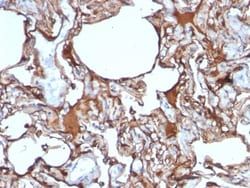
Antigen: PMEL17/SILV
Classification: Monoclonal
Concentration: 0.2mg/mL
Dilution: Flow Cytometry 5 - 10 ul/million cells in 0.1ml, Immunohistochemistry-Paraffin 1:100-1:200, Immunofluorescence 1:50 - 1:100
Gene Alias: D12S53EP1, gp100, ME20, ME20-M, melanocyte protein mel 17, Melanocyte protein Pmel 17, Melanocytes lineage-specific antigen GP100, Melanoma-associated ME20 antigen, melanosomal matrix protein17, PMEL17P100, premelanosome proteinME20M, SI, SIL, silver (mouse homolog) like, silver homolog (mouse), Silver locus protein homolog, silver, mouse, homolog of, SILVPmel17
Host Species: Mouse
Molecular Weight of Antigen: 95 kDa
Quantity: 0.02 mg
Research Discipline: __
Gene ID (Entrez): 6490
Target Species: Human, Canine (Negative), Rat (Negative)
Form: Purified
Applications: Flow Cytometry, Immunohistochemistry (Paraffin), Immunofluorescence
Clone: HMB45
Conjugate: Unconjugated
Formulation: 10mM PBS and 0.05% BSA with 0.05% Sodium Azide
Gene Symbols: PMEL
Immunogen: Extract of pigmented melanoma metastases from lymph nodes
Purification Method: Protein A or G purified
Regulatory Status: RUO
Primary or Secondary: Primary
Test Specificity: By immunohistochemistry, it specifically recognizes a protein in melanocytes and melanomas. This MAb reacts with junctional and blue nevus cells and variably with fetal and neonatal melanocytes. Intradermal nevi, normal adult melanocytes, and non-melanocytic cells are negative. It does not stain tumor cells of epithelial, lymphoid, glial, or mesenchymal origin. Metastatic amelanotic melanoma can often be confused with a variety of poorly differentiated carcinomas, large cell lymphomas, and sarcomas using H & E stains alone. It is also difficult to differentiate melanoma from spindle cell carcinomas and various types of mesenchymal neoplasms. This MAb stains fetal and neonatal melanocytes, junctional and blue nevus cells, and malignant melanoma. This MAb also stains Angiomyolipoma (PEComa).
Content And Storage: Store at 4C.
Isotype: IgG1 κ
Antigen: PMEL17/SILV
Classification: Monoclonal
Concentration: 0.2mg/mL
Dilution: Flow Cytometry 5 - 10 ul/million cells in 0.1ml, Immunohistochemistry-Paraffin 1:100-1:200, Immunofluorescence 1:50 - 1:100
Gene Alias: D12S53EP1, gp100, ME20, ME20-M, melanocyte protein mel 17, Melanocyte protein Pmel 17, Melanocytes lineage-specific antigen GP100, Melanoma-associated ME20 antigen, melanosomal matrix protein17, PMEL17P100, premelanosome proteinME20M, SI, SIL, silver (mouse homolog) like, silver homolog (mouse), Silver locus protein homolog, silver, mouse, homolog of, SILVPmel17
Host Species: Mouse
Molecular Weight of Antigen: 95 kDa
Quantity: 0.1 mg
Research Discipline: __
Gene ID (Entrez): 6490
Target Species: Human, Canine (Negative), Rat (Negative)
Form: Purified
Applications: Flow Cytometry, Immunohistochemistry (Paraffin), Immunofluorescence
Clone: HMB45
Conjugate: Unconjugated
Formulation: 10mM PBS and 0.05% BSA with 0.05% Sodium Azide
Gene Symbols: PMEL
Immunogen: Extract of pigmented melanoma metastases from lymph nodes
Purification Method: Protein A or G purified
Regulatory Status: RUO
Primary or Secondary: Primary
Test Specificity: By immunohistochemistry, it specifically recognizes a protein in melanocytes and melanomas. This MAb reacts with junctional and blue nevus cells and variably with fetal and neonatal melanocytes. Intradermal nevi, normal adult melanocytes, and non-melanocytic cells are negative. It does not stain tumor cells of epithelial, lymphoid, glial, or mesenchymal origin. Metastatic amelanotic melanoma can often be confused with a variety of poorly differentiated carcinomas, large cell lymphomas, and sarcomas using H & E stains alone. It is also difficult to differentiate melanoma from spindle cell carcinomas and various types of mesenchymal neoplasms. This MAb stains fetal and neonatal melanocytes, junctional and blue nevus cells, and malignant melanoma. This MAb also stains Angiomyolipoma (PEComa).
Content And Storage: Store at 4C.
Isotype: IgG1 κ