CD34 Antibody (SPM610), Novus Biologicals™
Manufacturer: Fischer Scientific
Select a Size
| Pack Size | SKU | Availability | Price |
|---|---|---|---|
| Each of 1 | NBP24457001-Each-of-1 | In Stock | ₹ 47,704.00 |
NBP24457001 - Each of 1
In Stock
Quantity
1
Base Price: ₹ 47,704.00
GST (18%): ₹ 8,586.72
Total Price: ₹ 56,290.72
Antigen
CD34
Classification
Monoclonal
Concentration
0.2mg/mL
Dilution
Western Blot 0.5 - 1.0 ug/ml, Flow Cytometry 0.5 - 1 ug/million cells in 0.1 ml, Immunohistochemistry-Paraffin 2 - 4 ug/ml, Immunofluorescence 0.5 - 1.0 ug/ml
Gene Alias
CD34 antigenhematopoietic progenitor cell antigen CD34, CD34 molecule
Host Species
Mouse
Purification Method
Protein A or G purified
Regulatory Status
RUO
Primary or Secondary
Primary
Test Specificity
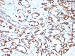
This antibody recognizes a carbohydrate epitope on a single chain, transmembrane, heavily glycosylated protein of 90-120kDa, which is identified as CD34 (VI international workshop on human differentiation antigens). Its expression is a hallmark for identifying pluripotent hematopoietic stem or progenitor cells. Its expression is gradually lost as lineage committed progenitors differentiate. CD34 is a marker of choice for staining blasts in acute myeloid leukemia. In addition, it is expressed by soft tissue tumors, such as solitary fibrous tumor and gastrointestinal stromal tumor. CD34 expression is also found in vascular endothelium. Additionally, proliferating endothelial cells overexpress this molecule than the non-proliferating endothelial cells. Anti-CD34 labels > 85% of angiosarcoma and Kaposi s sarcoma, but shows low specificity.
Content And Storage
Store at 4C.
Isotype
IgG1 κ
Applications
Western Blot, Flow Cytometry, Immunohistochemistry (Paraffin), Immunofluorescence
Clone
SPM610
Conjugate
Unconjugated
Formulation
10mM PBS and 0.05% BSA with 0.05% Sodium Azide
Gene Symbols
CD34
Immunogen
Recombinant full-length human HPCA1 protein
Quantity
0.1 mg
Research Discipline
Adaptive Immunity, Breast Cancer, Cancer, Cell Biology, Cellular Markers, Endothelial Cell Markers, Hematopoietic Stem Cell Markers, Hypoxia, Immunology, Innate Immunity, Mast Cell Markers, Mesenchymal Stem Cell Markers, Myeloid Cell Markers, Neuronal Cell Markers, Neuroscience, Stem Cell Markers, Tumor Suppressors
Gene ID (Entrez)
947
Target Species
Human, Rat
Form
Purified
Description
- Ensure accurate, reproducible results in Western Blot, Flow Cytometry, Immunohistochemistry (Paraffin), Immunofluorescence CD34 Monoclonal specifically detects CD34 in Human, Rat samples
- It is validated for Western Blot, Flow Cytometry, Immunohistochemistry, Immunocytochemistry/Immunofluorescence, Immunohistochemistry-Paraffin, Immunofluorescence.
Compare Similar Items
Show Difference

Antigen: CD34
Classification: Monoclonal
Concentration: 0.2mg/mL
Dilution: Western Blot 0.5 - 1.0 ug/ml, Flow Cytometry 0.5 - 1 ug/million cells in 0.1 ml, Immunohistochemistry-Paraffin 2 - 4 ug/ml, Immunofluorescence 0.5 - 1.0 ug/ml
Gene Alias: CD34 antigenhematopoietic progenitor cell antigen CD34, CD34 molecule
Host Species: Mouse
Purification Method: Protein A or G purified
Regulatory Status: RUO
Primary or Secondary: Primary
Test Specificity: This antibody recognizes a carbohydrate epitope on a single chain, transmembrane, heavily glycosylated protein of 90-120kDa, which is identified as CD34 (VI international workshop on human differentiation antigens). Its expression is a hallmark for identifying pluripotent hematopoietic stem or progenitor cells. Its expression is gradually lost as lineage committed progenitors differentiate. CD34 is a marker of choice for staining blasts in acute myeloid leukemia. In addition, it is expressed by soft tissue tumors, such as solitary fibrous tumor and gastrointestinal stromal tumor. CD34 expression is also found in vascular endothelium. Additionally, proliferating endothelial cells overexpress this molecule than the non-proliferating endothelial cells. Anti-CD34 labels > 85% of angiosarcoma and Kaposi s sarcoma, but shows low specificity.
Content And Storage: Store at 4C.
Isotype: IgG1 κ
Applications: Western Blot, Flow Cytometry, Immunohistochemistry (Paraffin), Immunofluorescence
Clone: SPM610
Conjugate: Unconjugated
Formulation: 10mM PBS and 0.05% BSA with 0.05% Sodium Azide
Gene Symbols: CD34
Immunogen: Recombinant full-length human HPCA1 protein
Quantity: 0.1 mg
Research Discipline: Adaptive Immunity, Breast Cancer, Cancer, Cell Biology, Cellular Markers, Endothelial Cell Markers, Hematopoietic Stem Cell Markers, Hypoxia, Immunology, Innate Immunity, Mast Cell Markers, Mesenchymal Stem Cell Markers, Myeloid Cell Markers, Neuronal Cell Markers, Neuroscience, Stem Cell Markers, Tumor Suppressors
Gene ID (Entrez): 947
Target Species: Human, Rat
Form: Purified
Antigen: CD34
Classification: Monoclonal
Concentration: 0.2mg/mL
Dilution: Western Blot 0.5 - 1.0 ug/ml, Flow Cytometry 0.5 - 1 ug/million cells in 0.1 ml, Immunohistochemistry-Paraffin 2 - 4 ug/ml, Immunofluorescence 0.5 - 1.0 ug/ml
Gene Alias: CD34 antigenhematopoietic progenitor cell antigen CD34, CD34 molecule
Host Species: Mouse
Purification Method: Protein A or G purified
Regulatory Status: RUO
Primary or Secondary: Primary
Test Specificity: This antibody recognizes a carbohydrate epitope on a single chain, transmembrane, heavily glycosylated protein of 90-120kDa, which is identified as CD34 (VI international workshop on human differentiation antigens). Its expression is a hallmark for identifying pluripotent hematopoietic stem or progenitor cells. Its expression is gradually lost as lineage committed progenitors differentiate. CD34 is a marker of choice for staining blasts in acute myeloid leukemia. In addition, it is expressed by soft tissue tumors, such as solitary fibrous tumor and gastrointestinal stromal tumor. CD34 expression is also found in vascular endothelium. Additionally, proliferating endothelial cells overexpress this molecule than the non-proliferating endothelial cells. Anti-CD34 labels > 85% of angiosarcoma and Kaposi s sarcoma, but shows low specificity.
Content And Storage: Store at 4C.
Isotype: IgG1 κ
Applications: Western Blot, Flow Cytometry, Immunohistochemistry (Paraffin), Immunofluorescence
Clone: SPM610
Conjugate: Unconjugated
Formulation: 10mM PBS and 0.05% BSA with 0.05% Sodium Azide
Gene Symbols: CD34
Immunogen: Recombinant full-length human HPCA1 protein
Quantity: 0.2 mg
Research Discipline: Adaptive Immunity, Breast Cancer, Cancer, Cell Biology, Cellular Markers, Endothelial Cell Markers, Hematopoietic Stem Cell Markers, Hypoxia, Immunology, Innate Immunity, Mast Cell Markers, Mesenchymal Stem Cell Markers, Myeloid Cell Markers, Neuronal Cell Markers, Neuroscience, Stem Cell Markers, Tumor Suppressors
Gene ID (Entrez): 947
Target Species: Human, Rat
Form: Purified

Antigen: DPPIV/CD26
Classification: Monoclonal
Concentration: 0.2 mg/mL
Dilution: Flow Cytometry 0.5 - 1 ug/million cells in 0.1 ml, Immunohistochemistry-Frozen 0.5 - 1.0 ug/ml, Immunofluorescence 0.5 - 1.0 ug/ml
Gene Alias: ADABP, ADCP-2, ADCP2DPP IV, Adenosine deaminase complexing protein 2TP103, CD26 antigen, CD26T-cell activation antigen CD26, dipeptidyl peptidase 4, Dipeptidyl peptidase IV, dipeptidylpeptidase 4, dipeptidyl-peptidase 4, dipeptidylpeptidase IV (CD26, adenosine deaminase complexing protein 2), DPPIV, EC 3.4.14.5
Host Species: Mouse
Purification Method: Protein A or G purified
Regulatory Status: RUO
Primary or Secondary: Primary
Test Specificity: Recognizes a glycoprotein of 110kDa, identified as CD26 (Workshop VI; Code: N-L039). It is an atypical serine protease belonging to the prolyl oligopeptidase family. It is expressed on lymphocyte cells and is upregulated during T-cell activation. CD26 is also expressed on activated B cells and natural killer cells and abundantly on epithelia. CD26 is implicated in a variety of biological functions including T-cell activation, cell adhesion with extracellular matrix such as fibronectin or collagens, and in HIV infection. Cross-linking of CD26 using this antibody dramatically enhances the anti-CD3-induced IL-2 production. In Western blotting, this MAb reacts with only glycosylated CD26, but not with the deglycosylated form. It does not prevent ADA binding to CD26.
Content And Storage: Store at 4C.
Isotype: IgG2b κ
Applications: Flow Cytometry, Immunohistochemistry (Frozen), Immunofluorescence
Clone: 202.36
Conjugate: Unconjugated
Formulation: 10mM PBS and 0.05% BSA with 0.05% Sodium Azide
Gene Symbols: DPP4
Immunogen: Human T cell clone
Quantity: 0.02 mg
Research Discipline: Adaptive Immunity, Cellular Markers, GPCR, Immunology
Gene ID (Entrez): 1803
Target Species: Human, Rat, Porcine (Negative), Sheep (Negative)
Form: Purified