WT1 Antibody (WT1/857), Novus Biologicals™
Manufacturer: Fischer Scientific
Select a Size
| Pack Size | SKU | Availability | Price |
|---|---|---|---|
| Each of 1 | NBP24460601-Each-of-1 | In Stock | ₹ 46,636.00 |
NBP24460601 - Each of 1
In Stock
Quantity
1
Base Price: ₹ 46,636.00
GST (18%): ₹ 8,394.48
Total Price: ₹ 55,030.48
Antigen
WT1
Classification
Monoclonal
Concentration
0.2mg/mL
Dilution
Flow Cytometry 0.5 - 1 ug/million cells in 0.1 ml, Immunohistochemistry-Paraffin 0.5 - 1.0 ug/ml, Immunofluorescence 0.5 - 1.0 ug/ml
Gene Accession No.
P19544
Gene Symbols
WT1
Immunogen
Recombinant human WT1 protein
Quantity
0.1 mg
Research Discipline
Angiogenesis, Cancer, Cell Cycle and Replication, Cellular Markers, Chromatin Research, Tumor Suppressors
Gene ID (Entrez)
7490
Target Species
Human, Mouse, Rat
Form
Purified
Applications
Flow Cytometry, Immunohistochemistry (Paraffin), Immunofluorescence
Clone
WT1/857
Conjugate
Unconjugated
Formulation
1.0mM PBS and 0.05% BSA with 0.05% Sodium Azide
Gene Alias
AWT1, NPHS4GUD, WAGR, Wilms tumor 1, Wilms tumor protein, WIT-2, WT33
Host Species
Mouse
Purification Method
Protein A or G purified
Regulatory Status
RUO
Primary or Secondary
Primary
Test Specificity
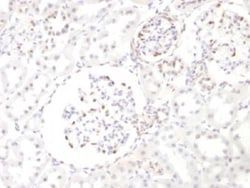
Recognizes a 47-55kDa-tumor suppressor protein, identified as Wilm's Tumor (WT1) protein. The antibody reacts with all isoforms of the full-length WT1 and also identifies WT1 lacking exon 2-encoded amino acids, frequently found in subsets of sporadic Wilm s tumors.WT1, a sporadic and familial pediatric kidney tumor, is genetically heterogeneous. Wilm s tumor is associated with mutations of WT1, a zinc-finger transcription factor that is essential for the development of the metanephric kidney and the urogenital system. The WT1 gene is normally expressed in fetal kidney and mesothelium, and its expression has been suggested as a marker for Wilm s tumor and mesothelioma. WT1 protein has been identified in proliferative mesothelial cells, malignant mesothelioma, ovarian carcinoma, gonadoblastoma, nephroblastoma, and desmoplastic small round cell tumor. Lung adenocarcinomas rarely stain positive with this antibody. WT1 protein expression in mesothelial cells has become a reliable marker for
Content And Storage
Store at 4C.
Isotype
IgG1 κ
Description
- Ensure accurate, reproducible results in Flow Cytometry, Immunohistochemistry (Paraffin), Immunofluorescence WT1 Monoclonal specifically detects WT1 in Human, Rat samples
- It is validated for Immunohistochemistry, Immunohistochemistry-Paraffin.
Compare Similar Items
Show Difference

Antigen: WT1
Classification: Monoclonal
Concentration: 0.2mg/mL
Dilution: Flow Cytometry 0.5 - 1 ug/million cells in 0.1 ml, Immunohistochemistry-Paraffin 0.5 - 1.0 ug/ml, Immunofluorescence 0.5 - 1.0 ug/ml
Gene Accession No.: P19544
Gene Symbols: WT1
Immunogen: Recombinant human WT1 protein
Quantity: 0.1 mg
Research Discipline: Angiogenesis, Cancer, Cell Cycle and Replication, Cellular Markers, Chromatin Research, Tumor Suppressors
Gene ID (Entrez): 7490
Target Species: Human, Mouse, Rat
Form: Purified
Applications: Flow Cytometry, Immunohistochemistry (Paraffin), Immunofluorescence
Clone: WT1/857
Conjugate: Unconjugated
Formulation: 1.0mM PBS and 0.05% BSA with 0.05% Sodium Azide
Gene Alias: AWT1, NPHS4GUD, WAGR, Wilms tumor 1, Wilms tumor protein, WIT-2, WT33
Host Species: Mouse
Purification Method: Protein A or G purified
Regulatory Status: RUO
Primary or Secondary: Primary
Test Specificity: Recognizes a 47-55kDa-tumor suppressor protein, identified as Wilm's Tumor (WT1) protein. The antibody reacts with all isoforms of the full-length WT1 and also identifies WT1 lacking exon 2-encoded amino acids, frequently found in subsets of sporadic Wilm s tumors.WT1, a sporadic and familial pediatric kidney tumor, is genetically heterogeneous. Wilm s tumor is associated with mutations of WT1, a zinc-finger transcription factor that is essential for the development of the metanephric kidney and the urogenital system. The WT1 gene is normally expressed in fetal kidney and mesothelium, and its expression has been suggested as a marker for Wilm s tumor and mesothelioma. WT1 protein has been identified in proliferative mesothelial cells, malignant mesothelioma, ovarian carcinoma, gonadoblastoma, nephroblastoma, and desmoplastic small round cell tumor. Lung adenocarcinomas rarely stain positive with this antibody. WT1 protein expression in mesothelial cells has become a reliable marker for
Content And Storage: Store at 4C.
Isotype: IgG1 κ
Antigen: WT1
Classification: Monoclonal
Concentration: 0.2mg/mL
Dilution: Flow Cytometry 0.5 - 1 ug/million cells in 0.1 ml, Immunohistochemistry-Paraffin 0.5 - 1.0 ug/ml, Immunofluorescence 0.5 - 1.0 ug/ml
Gene Accession No.: P19544
Gene Symbols: WT1
Immunogen: Recombinant human WT1 protein
Quantity: 0.2 mg
Research Discipline: Angiogenesis, Cancer, Cell Cycle and Replication, Cellular Markers, Chromatin Research, Tumor Suppressors
Gene ID (Entrez): 7490
Target Species: Human, Mouse, Rat
Form: Purified
Applications: Flow Cytometry, Immunohistochemistry (Paraffin), Immunofluorescence
Clone: WT1/857
Conjugate: Unconjugated
Formulation: 1.0mM PBS and 0.05% BSA with 0.05% Sodium Azide
Gene Alias: AWT1, NPHS4GUD, WAGR, Wilms tumor 1, Wilms tumor protein, WIT-2, WT33
Host Species: Mouse
Purification Method: Protein A or G purified
Regulatory Status: RUO
Primary or Secondary: Primary
Test Specificity: Recognizes a 47-55kDa-tumor suppressor protein, identified as Wilm's Tumor (WT1) protein. The antibody reacts with all isoforms of the full-length WT1 and also identifies WT1 lacking exon 2-encoded amino acids, frequently found in subsets of sporadic Wilm s tumors.WT1, a sporadic and familial pediatric kidney tumor, is genetically heterogeneous. Wilm s tumor is associated with mutations of WT1, a zinc-finger transcription factor that is essential for the development of the metanephric kidney and the urogenital system. The WT1 gene is normally expressed in fetal kidney and mesothelium, and its expression has been suggested as a marker for Wilm s tumor and mesothelioma. WT1 protein has been identified in proliferative mesothelial cells, malignant mesothelioma, ovarian carcinoma, gonadoblastoma, nephroblastoma, and desmoplastic small round cell tumor. Lung adenocarcinomas rarely stain positive with this antibody. WT1 protein expression in mesothelial cells has become a reliable marker for
Content And Storage: Store at 4C.
Isotype: IgG1 κ
Antigen: CD53
Classification: Monoclonal
Concentration: 0.2 mg/mL
Dilution: Flow Cytometry 0.5 - 1 ug/million cells in 0.1 ml, SDS-Page, Immunofluorescence 0.5 - 1.0 ug/ml
Gene Accession No.: __
Gene Symbols: CD53
Immunogen: Human Sezary cells
Quantity: 0.02 mg
Research Discipline: Immunology
Gene ID (Entrez): 963
Target Species: Human
Form: Purified
Applications: Flow Cytometry, SDS-Page, Immunofluorescence
Clone: 63-5A3
Conjugate: Unconjugated
Formulation: 10mM PBS and 0.05% BSA with 0.05% Sodium Azide
Gene Alias: antigen MOX44 identified by monoclonal MRC-OX44, CD53 antigentetraspanin-25, CD53 glycoprotein, CD53 molecule, CD53 tetraspan antigen, cell surface antigen, Cell surface glycoprotein CD53, MOX44transmembrane glycoprotein, Tetraspanin-25, tspan-25, TSPAN25leukocyte surface antigen CD53
Host Species: Mouse
Purification Method: Protein A or G purified
Regulatory Status: RUO
Primary or Secondary: Primary
Test Specificity: Recognizes a protein of 33-55kDa, identified as CD53 (Workshop V; Code CD53.1). CD53 is expressed on monocytes, and macrophages, granulocytes, dendritic cells, osteoblasts and osteoclasts, NK cells, and on T- and B-cells from every stage of differentiation but is absent from platelets, erythrocytes, and non-haemopoietic cells. CD53 is a member of a family of tetraspan transmembrane proteins, including CD9, CD37, CD63, CD81, and CD82. It associates with integrins, MHC class II molecules, and a tyrosine phosphatase and plays a role in cellular activation as part of a signal transduction complex involving other membrane glycoproteins. Defects of CD53 expression on neutrophils appear to be related with recurrent infectious diseases. Cross-linking CD53 using CD53 antibodies led to cytoplasmic calcium fluxes in B cells, monocytes, and granulocytes and activation of the monocyte oxidative burst.
Content And Storage: Store at 4C.
Isotype: IgG2b κ