ABO, Blood Group A Antigen Antibody (3-3A), Novus Biologicals™
Manufacturer: Fischer Scientific
The price for this product is unavailable. Please request a quote
Antigen
ABO, Blood Group A Antigen
Classification
Monoclonal
Concentration
0.2mg/mL
Dilution
Immunohistochemistry-Paraffin 0.5 - 1.0 ug/ml, SDS-Page, Immunofluorescence 0.5 - 1.0 ug/ml
Gene Accession No.
P16442
Gene Symbols
ABO
Immunogen
Mucin isolated from an ovarian cyst fluid
Quantity
0.2 mg
Primary or Secondary
Primary
Test Specificity
This MAb preferably reacts with determinants of chain A and H type 3(Gal1-3GalNAc-R) and 4 (Gal1-3GalNAc-R), but not with type 1 and 2 chain structures. It is not reactive with immuno-dominant A trisaccharide. This MAb is applicable for tissue staining in tumor patients with blood groups A and AB. It shows a highly heterogeneous reactivity in human colon tumor tissue and adjacent mucosa. Blood-group antigens are generally defined as molecules formed by sequential addition of saccharides to the carbohydrate side chains of lipids and proteins detected on erythrocytes and certain epithelial cells. The A, B and H antigens are reported to undergo modulation during malignant cellular transformation. Blood group related antigens represent a group of carbohydrate determinants carried on both glycolipids and glycoproteins. They are usually mucin-type, and are detected on erythrocytes, certain epithelial cells, and in secretions of certain individuals. Sixteen genetically and biosynthetically di
Content And Storage
Store at 4C.
Isotype
IgG1 κ
Applications
Immunohistochemistry (Paraffin), SDS-Page, Immunofluorescence
Clone
3-3A
Conjugate
Unconjugated
Formulation
10mM PBS and 0.05% BSA with 0.05% Sodium Azide
Gene Alias
A transferase, A3GALNT, A3GALT1, ABO blood group (transferase A, alpha 1-3-N-acetylgalactosaminyltransferase;transferase B, alpha 1-3-galactosyltransferase), ABO glycosyltransferase, B transferase, B(A) alpha-1,3-galactosyltransferase, EC 2.4.1.37, EC 2.4.1.40, Fucosylglycoprotein 3-alpha-galactosyltransferase, Fucosylglycoprotein alpha-N-acetylgalactosaminyltransferase, Glycoprotein-fucosylgalactoside alpha-galactosyltransferase, Glycoprotein-fucosylgalactoside alpha-N-acetylgalactosaminyltransferase, GTB, Histo-blood group A transferase, histo-blood group A2 transferase, histo-blood group ABO system transferase, Histo-blood group B transferase, NAGAT
Host Species
Mouse
Purification Method
Protein A or G purified
Regulatory Status
RUO
Gene ID (Entrez)
28
Target Species
Human
Form
Purified
Related Products


Description
- Ensure accurate, reproducible results in Immunohistochemistry (Paraffin), Immunofluorescence ABO, Blood Group A Antigen Monoclonal specifically detects ABO, Blood Group A Antigen in Human samples
- It is validated for Immunohistochemistry, Immunohistochemistry-Paraffin.
Compare Similar Items
Show Difference
Antigen: ABO, Blood Group A Antigen
Classification: Monoclonal
Concentration: 0.2mg/mL
Dilution: Immunohistochemistry-Paraffin 0.5 - 1.0 ug/ml, SDS-Page, Immunofluorescence 0.5 - 1.0 ug/ml
Gene Accession No.: P16442
Gene Symbols: ABO
Immunogen: Mucin isolated from an ovarian cyst fluid
Quantity: 0.2 mg
Primary or Secondary: Primary
Test Specificity: This MAb preferably reacts with determinants of chain A and H type 3(Gal1-3GalNAc-R) and 4 (Gal1-3GalNAc-R), but not with type 1 and 2 chain structures. It is not reactive with immuno-dominant A trisaccharide. This MAb is applicable for tissue staining in tumor patients with blood groups A and AB. It shows a highly heterogeneous reactivity in human colon tumor tissue and adjacent mucosa. Blood-group antigens are generally defined as molecules formed by sequential addition of saccharides to the carbohydrate side chains of lipids and proteins detected on erythrocytes and certain epithelial cells. The A, B and H antigens are reported to undergo modulation during malignant cellular transformation. Blood group related antigens represent a group of carbohydrate determinants carried on both glycolipids and glycoproteins. They are usually mucin-type, and are detected on erythrocytes, certain epithelial cells, and in secretions of certain individuals. Sixteen genetically and biosynthetically di
Content And Storage: Store at 4C.
Isotype: IgG1 κ
Applications: Immunohistochemistry (Paraffin), SDS-Page, Immunofluorescence
Clone: 3-3A
Conjugate: Unconjugated
Formulation: 10mM PBS and 0.05% BSA with 0.05% Sodium Azide
Gene Alias: A transferase, A3GALNT, A3GALT1, ABO blood group (transferase A, alpha 1-3-N-acetylgalactosaminyltransferase;transferase B, alpha 1-3-galactosyltransferase), ABO glycosyltransferase, B transferase, B(A) alpha-1,3-galactosyltransferase, EC 2.4.1.37, EC 2.4.1.40, Fucosylglycoprotein 3-alpha-galactosyltransferase, Fucosylglycoprotein alpha-N-acetylgalactosaminyltransferase, Glycoprotein-fucosylgalactoside alpha-galactosyltransferase, Glycoprotein-fucosylgalactoside alpha-N-acetylgalactosaminyltransferase, GTB, Histo-blood group A transferase, histo-blood group A2 transferase, histo-blood group ABO system transferase, Histo-blood group B transferase, NAGAT
Host Species: Mouse
Purification Method: Protein A or G purified
Regulatory Status: RUO
Gene ID (Entrez): 28
Target Species: Human
Form: Purified
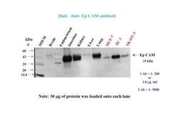
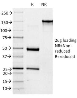
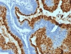
Antigen: EpCAM/TROP1
Classification: Monoclonal
Concentration: 0.2mg/mL
Dilution: Western Blot 0.5 - 1.0 ug/ml, Flow Cytometry 0.5 - 1 ug/million cells in 0.1 ml, Immunohistochemistry-Paraffin 0.5 - 1.0 ug/ml, Immunofluorescence 1 - 2 ug/ml
Gene Accession No.: P16422
Gene Symbols: EPCAM
Immunogen: A recombinant human EpCAM fragment from the cytoplasmic domain (around aa 280-350) (exact sequence is proprietary)
Quantity: 0.02 mg
Primary or Secondary: Primary
Test Specificity: EGP40 is a 40-43kDa transmembrane epithelial glycoprotein, also identified as epithelial specific antigen (ESA), or epithelial cellular adhesion molecule (Ep-CAM). It is expressed on baso-lateral cell surface in most simple epithelia and a vast majority of carcinomas. This antibody has been used to distinguish adenocarcinoma from pleural mesothelioma and hepatocellular carcinoma. This antibody is also useful in distinguishing serous carcinomas of the ovary from mesothelioma.
Content And Storage: Store at 4C.
Isotype: IgG2b κ
Applications: Western Blot, Flow Cytometry, Immunohistochemistry (Paraffin), Immunofluorescence
Clone: EGP40/1110
Conjugate: Unconjugated
Formulation: 10mM PBS and 0.05% BSA with 0.05% Sodium Azide
Gene Alias: 17-1A, 323/A3, ACSTD1, antigen identified by monoclonal AUA1, CD326 antigen, Cell surface glycoprotein Trop-1, chromosome 4, surface marker (35kD glycoprotein), DIAR5, EGP, EGP-2, EGP314, EGP40, EpCAM, epithelial cell adhesion molecule, Epithelial cell surface antigen, Epithelial glycoprotein, Epithelial glycoprotein 314, ESA, GA733-2EGP34, hEGP314, HNPCC8, KS 1/4 antigen, KS1/4, KSAHEA125, M1S2, M4S1Ly74, Major gastrointestinal tumor-associated protein GA733-2, MIC18MH99, MOC31, TACST-1, TACSTD1, TROP1CD326, Tumor-associated calcium signal transducer 1CO-17A
Host Species: Mouse
Purification Method: Protein A or G purified
Regulatory Status: RUO
Gene ID (Entrez): 4072
Target Species: Human, Mouse, Rat
Form: Purified
Antigen: EpCAM/TROP1
Classification: Monoclonal
Concentration: 0.2mg/mL
Dilution: Western Blot 0.5 - 1.0 ug/ml, Flow Cytometry 0.5 - 1 ug/million cells in 0.1 ml, Immunohistochemistry-Paraffin 0.5 - 1.0 ug/ml, Immunofluorescence 1 - 2 ug/ml
Gene Accession No.: P16422
Gene Symbols: EPCAM
Immunogen: A recombinant human EpCAM fragment from the cytoplasmic domain (around aa 280-350) (exact sequence is proprietary)
Quantity: 0.1 mg
Primary or Secondary: Primary
Test Specificity: EGP40 is a 40-43kDa transmembrane epithelial glycoprotein, also identified as epithelial specific antigen (ESA), or epithelial cellular adhesion molecule (Ep-CAM). It is expressed on baso-lateral cell surface in most simple epithelia and a vast majority of carcinomas. This antibody has been used to distinguish adenocarcinoma from pleural mesothelioma and hepatocellular carcinoma. This antibody is also useful in distinguishing serous carcinomas of the ovary from mesothelioma.
Content And Storage: Store at 4C.
Isotype: IgG2b κ
Applications: Western Blot, Flow Cytometry, Immunohistochemistry (Paraffin), Immunofluorescence
Clone: EGP40/1110
Conjugate: Unconjugated
Formulation: 10mM PBS and 0.05% BSA with 0.05% Sodium Azide
Gene Alias: 17-1A, 323/A3, ACSTD1, antigen identified by monoclonal AUA1, CD326 antigen, Cell surface glycoprotein Trop-1, chromosome 4, surface marker (35kD glycoprotein), DIAR5, EGP, EGP-2, EGP314, EGP40, EpCAM, epithelial cell adhesion molecule, Epithelial cell surface antigen, Epithelial glycoprotein, Epithelial glycoprotein 314, ESA, GA733-2EGP34, hEGP314, HNPCC8, KS 1/4 antigen, KS1/4, KSAHEA125, M1S2, M4S1Ly74, Major gastrointestinal tumor-associated protein GA733-2, MIC18MH99, MOC31, TACST-1, TACSTD1, TROP1CD326, Tumor-associated calcium signal transducer 1CO-17A
Host Species: Mouse
Purification Method: Protein A or G purified
Regulatory Status: RUO
Gene ID (Entrez): 4072
Target Species: Human, Mouse, Rat
Form: Purified