CD44 Antibody (HCAM/918), Novus Biologicals™
Manufacturer: Fischer Scientific
The price for this product is unavailable. Please request a quote
Antigen
CD44
Classification
Monoclonal
Concentration
0.2mg/mL
Dilution
Western Blot, Flow Cytometry 0.5 - 1 ug/million cells in 0.1 ml, Immunohistochemistry-Paraffin 0.25 - 0.5 ug/ml, Immunofluorescence 0.5 - 1.0 ug/ml
Gene Alias
CD44 antigen, CD44 molecule (Indian blood group), CD44R, CDw44, cell surface glycoprotein CD44, chondroitin sulfate proteoglycan 8, CSPG8, ECMR-III, Epican, Extracellular matrix receptor III, GP90 lymphocyte homing/adhesion receptor, HCELL, hematopoietic cell E- and L-selectin ligand, Heparan sulfate proteoglycan, Hermes antigen, homing function and Indian blood group system, HUTCH-I, Hyaluronate receptor, IN, LHR, MC56, MDU2CD44 antigen (homing function and Indian blood group system), MDU3CDW44, MIC4MGC10468, Pgp1, PGP-1, PGP-I, Phagocytic glycoprotein 1, Phagocytic glycoprotein I
Host Species
Mouse
Purification Method
Protein A or G purified
Regulatory Status
RUO
Primary or Secondary
Primary
Test Specificity
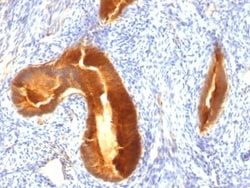
Recognizes a cell surface glycoprotein of 80-95kDa (CD44) on lymphocytes, monocytes, and granulocytes (Leucocyte Typing Workshop V). Its epitope is resistant to digestion by trypsin and chymotrypsin. The CD44 family of glycoproteins exists in a number of variant isoforms, the most common being the standard 85-95kDa or hematopoietic variant (CD44s). Higher molecular weight isoforms are described in epithelial cells (CD44v), which are believed to function in intercellular adhesion and stromal binding. CD44 immunostaining is commonly used for the discrimination of urothelial transitional cell carcinoma in-situ from non-neoplastic changes in the urothelium.
Content And Storage
Store at 4C.
Isotype
IgG2a κ
Applications
Western Blot, Flow Cytometry, Immunohistochemistry (Paraffin), Immunofluorescence
Clone
HCAM/918
Conjugate
Unconjugated
Formulation
10mM PBS and 0.05% BSA with 0.05% Sodium Azide
Gene Symbols
CD44
Immunogen
Recombinant human HCAM protein
Quantity
0.2 mg
Research Discipline
Cancer, Cell Biology, Extracellular Matrix, Hematopoietic Stem Cell Markers, Immunology, Mesenchymal Stem Cell Markers, Signal Transduction, Stem Cell Markers
Gene ID (Entrez)
960
Target Species
Human, Primate
Form
Purified
Description
- Ensure accurate, reproducible results in Flow Cytometry, Immunohistochemistry (Paraffin), Immunofluorescence CD44 Monoclonal specifically detects CD44 in Human, Chimpanzee, Baboon, Monkey samples
- It is validated for Western Blot, Flow Cytometry, Immunohistochemistry, Immunocytochemistry/Immunofluorescence, Immunohistochemistry-Paraffin, Functional.
Compare Similar Items
Show Difference
Antigen: CD44
Classification: Monoclonal
Concentration: 0.2mg/mL
Dilution: Western Blot, Flow Cytometry 0.5 - 1 ug/million cells in 0.1 ml, Immunohistochemistry-Paraffin 0.25 - 0.5 ug/ml, Immunofluorescence 0.5 - 1.0 ug/ml
Gene Alias: CD44 antigen, CD44 molecule (Indian blood group), CD44R, CDw44, cell surface glycoprotein CD44, chondroitin sulfate proteoglycan 8, CSPG8, ECMR-III, Epican, Extracellular matrix receptor III, GP90 lymphocyte homing/adhesion receptor, HCELL, hematopoietic cell E- and L-selectin ligand, Heparan sulfate proteoglycan, Hermes antigen, homing function and Indian blood group system, HUTCH-I, Hyaluronate receptor, IN, LHR, MC56, MDU2CD44 antigen (homing function and Indian blood group system), MDU3CDW44, MIC4MGC10468, Pgp1, PGP-1, PGP-I, Phagocytic glycoprotein 1, Phagocytic glycoprotein I
Host Species: Mouse
Purification Method: Protein A or G purified
Regulatory Status: RUO
Primary or Secondary: Primary
Test Specificity: Recognizes a cell surface glycoprotein of 80-95kDa (CD44) on lymphocytes, monocytes, and granulocytes (Leucocyte Typing Workshop V). Its epitope is resistant to digestion by trypsin and chymotrypsin. The CD44 family of glycoproteins exists in a number of variant isoforms, the most common being the standard 85-95kDa or hematopoietic variant (CD44s). Higher molecular weight isoforms are described in epithelial cells (CD44v), which are believed to function in intercellular adhesion and stromal binding. CD44 immunostaining is commonly used for the discrimination of urothelial transitional cell carcinoma in-situ from non-neoplastic changes in the urothelium.
Content And Storage: Store at 4C.
Isotype: IgG2a κ
Applications: Western Blot, Flow Cytometry, Immunohistochemistry (Paraffin), Immunofluorescence
Clone: HCAM/918
Conjugate: Unconjugated
Formulation: 10mM PBS and 0.05% BSA with 0.05% Sodium Azide
Gene Symbols: CD44
Immunogen: Recombinant human HCAM protein
Quantity: 0.2 mg
Research Discipline: Cancer, Cell Biology, Extracellular Matrix, Hematopoietic Stem Cell Markers, Immunology, Mesenchymal Stem Cell Markers, Signal Transduction, Stem Cell Markers
Gene ID (Entrez): 960
Target Species: Human, Primate
Form: Purified

Antigen: MUC-1
Classification: Monoclonal
Concentration: 0.2mg/mL
Dilution: Flow Cytometry 0.5 - 1 ug/million cells in 0.1 ml, Immunohistochemistry-Paraffin 0.25 - 0.5 ug/ml, Immunofluorescence 1 - 2 ug/ml
Gene Alias: Breast carcinoma-associated antigen DF3, Carcinoma-associated mucin, CD227, CD227 antigen, DF3 antigen, EMA, episialin, H23 antigen, H23AG, KL-6, MAM6, MUC-1, MUC1/ZD, mucin 1, cell surface associated, mucin 1, transmembrane, mucin-1, Peanut-reactive urinary mucin, PEMMUC-1/SEC, PEMT, Polymorphic epithelial mucin, PUMMUC-1/X, tumor associated epithelial mucin, Tumor-associated epithelial membrane antigen, Tumor-associated mucin
Host Species: Mouse
Purification Method: Protein A or G purified
Regulatory Status: RUO
Primary or Secondary: Primary
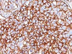
Test Specificity: This MAb reacts with MUC1. The dominant epitope of this MAb has not yet been determined. MUC1 is a large cell surface mucin glycoprotein expressed by most glandular and ductal epithelial cells and some hematopoietic cell lineages. It is expressed on most secretory epithelium, including mammary gland and some hematopoietic cells. It is expressed abundantly in lactating mammary glands and over expressed abundantly in >90% breast carcinomas and metastases. Transgenic MUC1 has been shown to associate with all four c-erbB receptors and localize with c-erbB1 (EGFR) in lactating glands. The MUC1 gene contains seven exons and produces several different alternatively spliced variants. The major expressed form of MUC1 uses all seven exons and is a type 1 transmembrane protein with a large extracellular tandem repeat domain. The tandem repeat domain is highly O glycosylated and alterations in glycosylation have been shown in epithelial cancer cells. Antibody to EMA is useful as a pan-epithelia
Content And Storage: Store at 4C.
Isotype: IgG1 κ
Applications: Flow Cytometry, Immunohistochemistry (Paraffin), Immunofluorescence
Clone: MUC1/955
Conjugate: Unconjugated
Formulation: 10mM PBS and 0.05% BSA with 0.05% Sodium Azide
Gene Symbols: MUC1
Immunogen: Human milk-fat globule membranes (HMFGM)
Quantity: 0.02 mg
Research Discipline: Cancer, Cellular Markers, Extracellular Matrix
Gene ID (Entrez): 4582
Target Species: Human, Mouse
Form: Purified
Antigen: MUC-1
Classification: Monoclonal
Concentration: 0.2mg/mL
Dilution: Flow Cytometry 0.5 - 1 ug/million cells in 0.1 ml, Immunohistochemistry-Paraffin 0.25 - 0.5 ug/ml, Immunofluorescence 1 - 2 ug/ml
Gene Alias: Breast carcinoma-associated antigen DF3, Carcinoma-associated mucin, CD227, CD227 antigen, DF3 antigen, EMA, episialin, H23 antigen, H23AG, KL-6, MAM6, MUC-1, MUC1/ZD, mucin 1, cell surface associated, mucin 1, transmembrane, mucin-1, Peanut-reactive urinary mucin, PEMMUC-1/SEC, PEMT, Polymorphic epithelial mucin, PUMMUC-1/X, tumor associated epithelial mucin, Tumor-associated epithelial membrane antigen, Tumor-associated mucin
Host Species: Mouse
Purification Method: Protein A or G purified
Regulatory Status: RUO
Primary or Secondary: Primary
Test Specificity: This MAb reacts with MUC1. The dominant epitope of this MAb has not yet been determined. MUC1 is a large cell surface mucin glycoprotein expressed by most glandular and ductal epithelial cells and some hematopoietic cell lineages. It is expressed on most secretory epithelium, including mammary gland and some hematopoietic cells. It is expressed abundantly in lactating mammary glands and over expressed abundantly in >90% breast carcinomas and metastases. Transgenic MUC1 has been shown to associate with all four c-erbB receptors and localize with c-erbB1 (EGFR) in lactating glands. The MUC1 gene contains seven exons and produces several different alternatively spliced variants. The major expressed form of MUC1 uses all seven exons and is a type 1 transmembrane protein with a large extracellular tandem repeat domain. The tandem repeat domain is highly O glycosylated and alterations in glycosylation have been shown in epithelial cancer cells. Antibody to EMA is useful as a pan-epithelia
Content And Storage: Store at 4C.
Isotype: IgG1 κ
Applications: Flow Cytometry, Immunohistochemistry (Paraffin), Immunofluorescence
Clone: MUC1/955
Conjugate: Unconjugated
Formulation: 10mM PBS and 0.05% BSA with 0.05% Sodium Azide
Gene Symbols: MUC1
Immunogen: Human milk-fat globule membranes (HMFGM)
Quantity: 0.1 mg
Research Discipline: Cancer, Cellular Markers, Extracellular Matrix
Gene ID (Entrez): 4582
Target Species: Human, Mouse
Form: Purified