Cytokeratin 10/13 Antibody (SPM262), Novus Biologicals™
Manufacturer: Fischer Scientific
Select a Size
| Pack Size | SKU | Availability | Price |
|---|---|---|---|
| Each of 1 | NBP24471001-Each-of-1 | In Stock | ₹ 46,636.00 |
NBP24471001 - Each of 1
In Stock
Quantity
1
Base Price: ₹ 46,636.00
GST (18%): ₹ 8,394.48
Total Price: ₹ 55,030.48
Antigen
Cytokeratin 10/13
Classification
Monoclonal
Concentration
0.2mg/mL
Dilution
Western Blot 0.25 - 0.5 ug/ml, Flow Cytometry 0.5 - 1 ug/million cells in 0.1 ml, Immunohistochemistry-Paraffin 0.5 - 1.0 ug/ml, Immunofluorescence 0.5 - 1.0 ug/ml
Gene Alias
BCIE, BIE, CK10, CK-10, cytokeratin 10, Cytokeratin-10, EHK, K10keratosis palmaris et plantaris, keratin 10, keratin, type I cytoskeletal 10, keratin-10, KPP
Host Species
Mouse
Purification Method
Protein A or G purified
Regulatory Status
RUO
Primary or Secondary
Primary
Test Specificity
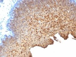
This antibody recognizes cytokeratin 10 (56.5kDa) and cytokeratin 13 (53kDa) in Western blotting. It recognizes only cytokeratin 13 in formalin-fixed, paraffin-embedded tissue sections. It does not react with cytokeratin 10 positive, cytokeratin 13 negative epithelia such as epidermis. However, on frozen sections this MAb serves as differentiation-related marker of all stratified epithelia; it stains all suprabasal cells in both cornifying and non-cornifying stratified epithelia and more differentiated cells of squamous carcinomas.
Content And Storage
Store at 4C.
Isotype
IgG2a κ
Applications
Western Blot, Flow Cytometry, Immunohistochemistry (Paraffin), Immunofluorescence
Clone
SPM262
Conjugate
Unconjugated
Formulation
10mM PBS and 0.05% BSA with 0.05% Sodium Azide
Gene Symbols
KRT10
Immunogen
Cytoskeletal preparation extracted from human ectocervical epithelium.
Quantity
0.1 mg
Research Discipline
Cytoskeleton Markers
Gene ID (Entrez)
3858
Target Species
Human, Feline
Form
Purified
Description
- Ensure accurate, reproducible results in Western Blot, Flow Cytometry, Immunohistochemistry (Paraffin), Immunofluorescence Cytokeratin 10/13 Monoclonal specifically detects Cytokeratin 10/13 in Human, Feline samples
- It is validated for Western Blot, Flow Cytometry, Immunohistochemistry, Immunocytochemistry/Immunofluorescence, Immunohistochemistry-Paraffin, Immunofluorescence.
Compare Similar Items
Show Difference

Antigen: Cytokeratin 10/13
Classification: Monoclonal
Concentration: 0.2mg/mL
Dilution: Western Blot 0.25 - 0.5 ug/ml, Flow Cytometry 0.5 - 1 ug/million cells in 0.1 ml, Immunohistochemistry-Paraffin 0.5 - 1.0 ug/ml, Immunofluorescence 0.5 - 1.0 ug/ml
Gene Alias: BCIE, BIE, CK10, CK-10, cytokeratin 10, Cytokeratin-10, EHK, K10keratosis palmaris et plantaris, keratin 10, keratin, type I cytoskeletal 10, keratin-10, KPP
Host Species: Mouse
Purification Method: Protein A or G purified
Regulatory Status: RUO
Primary or Secondary: Primary
Test Specificity: This antibody recognizes cytokeratin 10 (56.5kDa) and cytokeratin 13 (53kDa) in Western blotting. It recognizes only cytokeratin 13 in formalin-fixed, paraffin-embedded tissue sections. It does not react with cytokeratin 10 positive, cytokeratin 13 negative epithelia such as epidermis. However, on frozen sections this MAb serves as differentiation-related marker of all stratified epithelia; it stains all suprabasal cells in both cornifying and non-cornifying stratified epithelia and more differentiated cells of squamous carcinomas.
Content And Storage: Store at 4C.
Isotype: IgG2a κ
Applications: Western Blot, Flow Cytometry, Immunohistochemistry (Paraffin), Immunofluorescence
Clone: SPM262
Conjugate: Unconjugated
Formulation: 10mM PBS and 0.05% BSA with 0.05% Sodium Azide
Gene Symbols: KRT10
Immunogen: Cytoskeletal preparation extracted from human ectocervical epithelium.
Quantity: 0.1 mg
Research Discipline: Cytoskeleton Markers
Gene ID (Entrez): 3858
Target Species: Human, Feline
Form: Purified
Antigen: Cytokeratin 10/13
Classification: Monoclonal
Concentration: 0.2mg/mL
Dilution: Western Blot 0.25 - 0.5 ug/ml, Flow Cytometry 0.5 - 1 ug/million cells in 0.1 ml, Immunohistochemistry-Paraffin 0.5 - 1.0 ug/ml, Immunofluorescence 0.5 - 1.0 ug/ml
Gene Alias: BCIE, BIE, CK10, CK-10, cytokeratin 10, Cytokeratin-10, EHK, K10keratosis palmaris et plantaris, keratin 10, keratin, type I cytoskeletal 10, keratin-10, KPP
Host Species: Mouse
Purification Method: Protein A or G purified
Regulatory Status: RUO
Primary or Secondary: Primary
Test Specificity: This antibody recognizes cytokeratin 10 (56.5kDa) and cytokeratin 13 (53kDa) in Western blotting. It recognizes only cytokeratin 13 in formalin-fixed, paraffin-embedded tissue sections. It does not react with cytokeratin 10 positive, cytokeratin 13 negative epithelia such as epidermis. However, on frozen sections this MAb serves as differentiation-related marker of all stratified epithelia; it stains all suprabasal cells in both cornifying and non-cornifying stratified epithelia and more differentiated cells of squamous carcinomas.
Content And Storage: Store at 4C.
Isotype: IgG2a κ
Applications: Western Blot, Flow Cytometry, Immunohistochemistry (Paraffin), Immunofluorescence
Clone: SPM262
Conjugate: Unconjugated
Formulation: 10mM PBS and 0.05% BSA with 0.05% Sodium Azide
Gene Symbols: KRT10
Immunogen: Cytoskeletal preparation extracted from human ectocervical epithelium.
Quantity: 0.2 mg
Research Discipline: Cytoskeleton Markers
Gene ID (Entrez): 3858
Target Species: Human, Feline
Form: Purified
Antigen: NCAM-1/CD56
Classification: Monoclonal
Concentration: 0.2mg/mL
Dilution: Western Blot 0.5 - 1.0 ug/ml, Flow Cytometry 0.5 - 1 ug/million cells in 0.1 ml, Immunohistochemistry-Paraffin 0.5 - 1.0 ug/ml, SDS-Page, Immunofluorescence 1 - 2 ug/ml
Gene Alias: CD56, CD56/ NCAM-1, CD56 antigen, MSK39, N-CAM-1, NCAM-1, NCAMantigen recognized by monoclonal 5.1H11, neural cell adhesion molecule 1, neural cell adhesion molecule, NCAM
Host Species: Mouse
Purification Method: Protein A or G purified
Regulatory Status: RUO
Primary or Secondary: Primary
Test Specificity: This MAb reacts with an extracellular domain (close to transmembrane) of CD56/NCAM. Three isoforms of neural cell adhesion molecule (NCAM) are produced by differential splicing of the RNA transcript from a single gene. The 135kDa isoform is the basic molecule, which is glycosylated or sialylated to produce the mature species. Anti-CD56 recognizes two proteins of the neural cell adhesion molecule, the basic molecule expressed on most neuroectodermally derived tissues and neoplasms (e.g. retinoblastoma, medulloblastomas, astrocytomas, neuroblastomas, and small cell carcinomas). It is also expressed on some mesodermally derived tumors (rhabdomyosarcoma). Anti-CD56 plays an important role in the diagnosis of nodal and nasal NK/T-cell lymphomas.
Content And Storage: Store at 4C.
Isotype: IgG1 κ
Applications: Western Blot, Flow Cytometry, Immunohistochemistry (Paraffin), SDS-Page, Immunofluorescence
Clone: NCAM1/795
Conjugate: Unconjugated
Formulation: 10mM PBS and 0.05% BSA with 0.05% Sodium Azide
Gene Symbols: NCAM1
Immunogen: Recombinant human NCAM1 protein
Quantity: 0.02 mg
Research Discipline: Astrocyte Markers, Cellular Markers, Cytokine Research, Cytoskeleton Markers, Growth and Development, Hematopoietic Stem Cell Markers, Immunology, Innate Immunity, Membrane Vesicle Markers, Neuronal Cell Markers, Neuronal Stem Cell Markers, Neuroscience, Stem Cell Markers
Gene ID (Entrez): 4684
Target Species: Human
Form: Purified