CD20 Antibody (IGEL/773), Novus Biologicals™
Manufacturer: Fischer Scientific
The price for this product is unavailable. Please request a quote
Antigen
CD20
Classification
Monoclonal
Concentration
0.2mg/mL
Dilution
Flow Cytometry 0.5 - 1 ug/million cells in 0.1 ml, Immunohistochemistry-Paraffin 0.5 - 1.0 ug/ml, SDS-Page, Immunofluorescence 0.5 - 1.0 ug/ml
Gene Accession No.
P11836
Gene Symbols
MS4A1
Immunogen
Recombinant human MS4A1 protein
Purification Method
Protein A or G purified
Regulatory Status
RUO
Primary or Secondary
Primary
Test Specificity
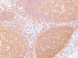
Recognizes a protein of 30-33kDa, which is identified as CD20. It is a non-Ig differentiation antigen of B-cells and its expression is restricted to normal and neoplastic B-cells, being absent from all other leukocytes and tissues. CD20 is expressed by pre B-cells and persists during all stages of B-cell maturation but is lost upon terminal differentiation into plasma cells. This MAb can be used for immunophenotyping of leukemia and malignant cells, B lymphocyte detection in peripheral blood and B cell localization in tissues. It reacts with the majority of B-cells present in peripheral blood and lymphoid tissues and their derived lymphomas. In lymphoid tissue, germinal center blasts and B-immunoblasts are particularly reactive. It is a reliable antibody for ascribing a B-cell phenotype in known lymphoid tissues. Rarely, CD20-positive T-cell lymphomas have been reported. Reactivity has also been noted with Reed-Sternberg cells in cases of Hodgkin s disease, particularly of lymphocyte p
Content And Storage
Store at 4C.
Isotype
IgG2a κ
Applications
Flow Cytometry, Immunohistochemistry (Paraffin), SDS-Page, Immunofluorescence
Clone
IGEL/773
Conjugate
Unconjugated
Formulation
10mM PBS and 0.05% BSA with 0.05% Sodium Azide
Gene Alias
B1, B-lymphocyte antigen CD20, B-lymphocyte cell-surface antigen B1, B-lymphocyte surface antigen B1, Bp35MGC3969, CD20 antigen, CD20 receptor, CD20S7, CVID5, LEU-16, Leukocyte surface antigen Leu-16, Membrane-spanning 4-domains subfamily A member 1, membrane-spanning 4-domains, subfamily A, member 1, MS4A2
Host Species
Mouse
Molecular Weight of Antigen
35 kDa
Quantity
0.2 mg
Research Discipline
Adaptive Immunity, Cancer Stem Cells, Cell Biology, Cytokine Research, Immunology, Signal Transduction, Stem Cell Markers, Tumor Biomarkers
Gene ID (Entrez)
931
Target Species
Human
Form
Purified
Description
- Ensure accurate, reproducible results in Flow Cytometry, Immunohistochemistry (Paraffin), Immunofluorescence CD20 Monoclonal specifically detects CD20 in Human samples
- It is validated for Western Blot, Flow Cytometry, Immunohistochemistry, Immunocytochemistry/Immunofluorescence, Immunohistochemistry-Paraffin, Multiplex Immunoassay.
Compare Similar Items
Show Difference
Antigen: CD20
Classification: Monoclonal
Concentration: 0.2mg/mL
Dilution: Flow Cytometry 0.5 - 1 ug/million cells in 0.1 ml, Immunohistochemistry-Paraffin 0.5 - 1.0 ug/ml, SDS-Page, Immunofluorescence 0.5 - 1.0 ug/ml
Gene Accession No.: P11836
Gene Symbols: MS4A1
Immunogen: Recombinant human MS4A1 protein
Purification Method: Protein A or G purified
Regulatory Status: RUO
Primary or Secondary: Primary
Test Specificity: Recognizes a protein of 30-33kDa, which is identified as CD20. It is a non-Ig differentiation antigen of B-cells and its expression is restricted to normal and neoplastic B-cells, being absent from all other leukocytes and tissues. CD20 is expressed by pre B-cells and persists during all stages of B-cell maturation but is lost upon terminal differentiation into plasma cells. This MAb can be used for immunophenotyping of leukemia and malignant cells, B lymphocyte detection in peripheral blood and B cell localization in tissues. It reacts with the majority of B-cells present in peripheral blood and lymphoid tissues and their derived lymphomas. In lymphoid tissue, germinal center blasts and B-immunoblasts are particularly reactive. It is a reliable antibody for ascribing a B-cell phenotype in known lymphoid tissues. Rarely, CD20-positive T-cell lymphomas have been reported. Reactivity has also been noted with Reed-Sternberg cells in cases of Hodgkin s disease, particularly of lymphocyte p
Content And Storage: Store at 4C.
Isotype: IgG2a κ
Applications: Flow Cytometry, Immunohistochemistry (Paraffin), SDS-Page, Immunofluorescence
Clone: IGEL/773
Conjugate: Unconjugated
Formulation: 10mM PBS and 0.05% BSA with 0.05% Sodium Azide
Gene Alias: B1, B-lymphocyte antigen CD20, B-lymphocyte cell-surface antigen B1, B-lymphocyte surface antigen B1, Bp35MGC3969, CD20 antigen, CD20 receptor, CD20S7, CVID5, LEU-16, Leukocyte surface antigen Leu-16, Membrane-spanning 4-domains subfamily A member 1, membrane-spanning 4-domains, subfamily A, member 1, MS4A2
Host Species: Mouse
Molecular Weight of Antigen: 35 kDa
Quantity: 0.2 mg
Research Discipline: Adaptive Immunity, Cancer Stem Cells, Cell Biology, Cytokine Research, Immunology, Signal Transduction, Stem Cell Markers, Tumor Biomarkers
Gene ID (Entrez): 931
Target Species: Human
Form: Purified

Antigen: CD20
Classification: Monoclonal
Concentration: 0.2 mg/mL
Dilution: Flow Cytometry 0.5 - 1 ug/million cells in 0.1 ml, Immunofluorescence 0.5 - 1.0 ug/ml
Gene Accession No.: P11836
Gene Symbols: MS4A1
Immunogen: Stimulated human leukocytes
Purification Method: Protein A or G purified
Regulatory Status: RUO
Primary or Secondary: Primary
Test Specificity: Recognizes a protein of 30-33kDa, which is identified as CD20 (Workshop V; Code CD20.12. Workshop IV; Code B17). It recognizes an extracellular domain of CD20. It is a non-Ig differentiation antigen of B-cells and its expression is restricted to normal and neoplastic B-cells, being absent from all other leukocytes and tissues. CD20 is expressed by pre B-cells and persists during all stages of B-cell maturation but is lost upon terminal differentiation into plasma cells. The protein passes through the membrane 4 times with both ends in cytoplasm and exposes one short and one longer loop to the external environment. CD20 is not glycosylated in resting B-cells and its cytoplasmic domains are differentially phosphorylated upon activation. It acts as calcium channel involved in B cell activation and cell cycle progression.
Content And Storage: Store at 4C.
Isotype: IgG3 κ
Applications: Flow Cytometry, Immunofluorescence
Clone: 109-3C2
Conjugate: Unconjugated
Formulation: 10mM PBS and 0.05% BSA with 0.05% Sodium Azide
Gene Alias: B1, B-lymphocyte antigen CD20, B-lymphocyte cell-surface antigen B1, B-lymphocyte surface antigen B1, Bp35MGC3969, CD20 antigen, CD20 receptor, CD20S7, CVID5, LEU-16, Leukocyte surface antigen Leu-16, Membrane-spanning 4-domains subfamily A member 1, membrane-spanning 4-domains, subfamily A, member 1, MS4A2
Host Species: Mouse
Molecular Weight of Antigen: 35 kDa
Quantity: 0.02 mg
Research Discipline: Adaptive Immunity, Cancer Stem Cells, Cell Biology, Cytokine Research, Immunology, Signal Transduction, Stem Cell Markers, Tumor Biomarkers
Gene ID (Entrez): 931
Target Species: Human
Form: Purified
Antigen: CD20
Classification: Monoclonal
Concentration: 0.2 mg/mL
Dilution: Flow Cytometry 0.5 - 1 ug/million cells in 0.1 ml, Immunofluorescence 0.5 - 1.0 ug/ml
Gene Accession No.: P11836
Gene Symbols: MS4A1
Immunogen: Stimulated human leukocytes
Purification Method: Protein A or G purified
Regulatory Status: RUO
Primary or Secondary: Primary
Test Specificity: Recognizes a protein of 30-33kDa, which is identified as CD20 (Workshop V; Code CD20.12. Workshop IV; Code B17). It recognizes an extracellular domain of CD20. It is a non-Ig differentiation antigen of B-cells and its expression is restricted to normal and neoplastic B-cells, being absent from all other leukocytes and tissues. CD20 is expressed by pre B-cells and persists during all stages of B-cell maturation but is lost upon terminal differentiation into plasma cells. The protein passes through the membrane 4 times with both ends in cytoplasm and exposes one short and one longer loop to the external environment. CD20 is not glycosylated in resting B-cells and its cytoplasmic domains are differentially phosphorylated upon activation. It acts as calcium channel involved in B cell activation and cell cycle progression.
Content And Storage: Store at 4C.
Isotype: IgG3 κ
Applications: Flow Cytometry, Immunofluorescence
Clone: 109-3C2
Conjugate: Unconjugated
Formulation: 10mM PBS and 0.05% BSA with 0.05% Sodium Azide
Gene Alias: B1, B-lymphocyte antigen CD20, B-lymphocyte cell-surface antigen B1, B-lymphocyte surface antigen B1, Bp35MGC3969, CD20 antigen, CD20 receptor, CD20S7, CVID5, LEU-16, Leukocyte surface antigen Leu-16, Membrane-spanning 4-domains subfamily A member 1, membrane-spanning 4-domains, subfamily A, member 1, MS4A2
Host Species: Mouse
Molecular Weight of Antigen: 35 kDa
Quantity: 0.1 mg
Research Discipline: Adaptive Immunity, Cancer Stem Cells, Cell Biology, Cytokine Research, Immunology, Signal Transduction, Stem Cell Markers, Tumor Biomarkers
Gene ID (Entrez): 931
Target Species: Human
Form: Purified
