CD63 Antibody (LAMP3/968), Novus Biologicals™
Manufacturer: Fischer Scientific
Select a Size
| Pack Size | SKU | Availability | Price |
|---|---|---|---|
| Each of 1 | NBP24481001-Each-of-1 | In Stock | ₹ 47,704.00 |
NBP24481001 - Each of 1
In Stock
Quantity
1
Base Price: ₹ 47,704.00
GST (18%): ₹ 8,586.72
Total Price: ₹ 56,290.72
Antigen
CD63
Classification
Monoclonal
Concentration
0.2mg/mL
Dilution
Flow Cytometry 0.5 - 1 ug/million cells in 0.1 ml, Immunohistochemistry-Paraffin 1 - 2 ug/ml, SDS-Page, Immunofluorescence 0.5 - 1.0 ug/ml
Gene Accession No.
P08962
Gene Symbols
CD63
Immunogen
Recombinant human full-length CD63 protein
Quantity
0.1 mg
Research Discipline
Autophagy, Cancer, Cardiovascular Biology, Cellular Signaling, Cytokine Research, Immunology, Innate Immunity, Lysosome Markers
Gene ID (Entrez)
967
Target Species
Human
Form
Purified
Applications
Flow Cytometry, Immunohistochemistry (Paraffin), SDS-Page, Immunofluorescence
Clone
LAMP3/968
Conjugate
Unconjugated
Formulation
10mM PBS and 0.05% BSA with 0.05% Sodium Azide
Gene Alias
CD63 antigen, CD63 antigen (melanoma 1 antigen), CD63 molecule, Granulophysin, LAMP-3, Lysosomal-associated membrane protein 3, ME491, melanoma 1 antigen, Melanoma-associated antigen ME491, melanoma-associated antigen MLA1, MLA1lysosome-associated membrane glycoprotein 3, Ocular melanoma-associated antigen, OMA81H, Tetraspanin-30, tspan-30, TSPAN30granulophysin
Host Species
Mouse
Purification Method
Protein A or G purified
Regulatory Status
RUO
Primary or Secondary
Primary
Test Specificity
This MAb recognizes protein of 26kDa-60kDa, which is identified as CD63. Its epitope is different from that of MAb LAMP3/529. The tetraspanins are integral membrane proteins expressed on cell surface and granular membranes of hematopoietic cells and are components of multi-molecular complexes with specific integrins. The tetraspanin CD63 is a lysosomal membrane glycoprotein that translocates to the plasma membrane after platelet activation. CD63 is expressed on activated platelets, monocytes and macrophages, and is weakly expressed on granulocytes, T cell and B cells. It is located on the basophilic granule membranes and on the plasma membranes of lymphocytes and granulocytes. CD63 is a member of the TM4 superfamily of leukocyte glycoproteins that includes CD9, CD37 and CD53, which contain four transmembrane regions. CD63 may play a role in phagocytic and intracellular lysosome-phagosome fusion events. CD63 deficiency is associated with Hermansky-Pudlak syndrome and is strongly express
Content And Storage
Store at 4C.
Isotype
IgG2a κ
Related Products


Description
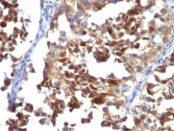
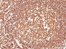
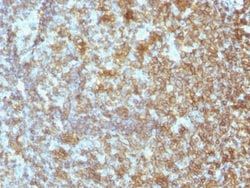
- Ensure accurate, reproducible results in Flow Cytometry, Immunohistochemistry (Paraffin), Immunofluorescence CD63 Monoclonal specifically detects CD63 in Human samples
- It is validated for Flow Cytometry, Immunohistochemistry, Immunohistochemistry-Paraffin, Flow (Intracellular).
Compare Similar Items
Show Difference
Antigen: CD63
Classification: Monoclonal
Concentration: 0.2mg/mL
Dilution: Flow Cytometry 0.5 - 1 ug/million cells in 0.1 ml, Immunohistochemistry-Paraffin 1 - 2 ug/ml, SDS-Page, Immunofluorescence 0.5 - 1.0 ug/ml
Gene Accession No.: P08962
Gene Symbols: CD63
Immunogen: Recombinant human full-length CD63 protein
Quantity: 0.1 mg
Research Discipline: Autophagy, Cancer, Cardiovascular Biology, Cellular Signaling, Cytokine Research, Immunology, Innate Immunity, Lysosome Markers
Gene ID (Entrez): 967
Target Species: Human
Form: Purified
Applications: Flow Cytometry, Immunohistochemistry (Paraffin), SDS-Page, Immunofluorescence
Clone: LAMP3/968
Conjugate: Unconjugated
Formulation: 10mM PBS and 0.05% BSA with 0.05% Sodium Azide
Gene Alias: CD63 antigen, CD63 antigen (melanoma 1 antigen), CD63 molecule, Granulophysin, LAMP-3, Lysosomal-associated membrane protein 3, ME491, melanoma 1 antigen, Melanoma-associated antigen ME491, melanoma-associated antigen MLA1, MLA1lysosome-associated membrane glycoprotein 3, Ocular melanoma-associated antigen, OMA81H, Tetraspanin-30, tspan-30, TSPAN30granulophysin
Host Species: Mouse
Purification Method: Protein A or G purified
Regulatory Status: RUO
Primary or Secondary: Primary
Test Specificity: This MAb recognizes protein of 26kDa-60kDa, which is identified as CD63. Its epitope is different from that of MAb LAMP3/529. The tetraspanins are integral membrane proteins expressed on cell surface and granular membranes of hematopoietic cells and are components of multi-molecular complexes with specific integrins. The tetraspanin CD63 is a lysosomal membrane glycoprotein that translocates to the plasma membrane after platelet activation. CD63 is expressed on activated platelets, monocytes and macrophages, and is weakly expressed on granulocytes, T cell and B cells. It is located on the basophilic granule membranes and on the plasma membranes of lymphocytes and granulocytes. CD63 is a member of the TM4 superfamily of leukocyte glycoproteins that includes CD9, CD37 and CD53, which contain four transmembrane regions. CD63 may play a role in phagocytic and intracellular lysosome-phagosome fusion events. CD63 deficiency is associated with Hermansky-Pudlak syndrome and is strongly express
Content And Storage: Store at 4C.
Isotype: IgG2a κ
Antigen: CD63
Classification: Monoclonal
Concentration: 0.2mg/mL
Dilution: Flow Cytometry 0.5 - 1 ug/million cells in 0.1 ml, Immunohistochemistry-Paraffin 1 - 2 ug/ml, SDS-Page, Immunofluorescence 0.5 - 1.0 ug/ml
Gene Accession No.: P08962
Gene Symbols: CD63
Immunogen: Recombinant human full-length CD63 protein
Quantity: 0.2 mg
Research Discipline: Autophagy, Cancer, Cardiovascular Biology, Cellular Signaling, Cytokine Research, Immunology, Innate Immunity, Lysosome Markers
Gene ID (Entrez): 967
Target Species: Human
Form: Purified
Applications: Flow Cytometry, Immunohistochemistry (Paraffin), SDS-Page, Immunofluorescence
Clone: LAMP3/968
Conjugate: Unconjugated
Formulation: 10mM PBS and 0.05% BSA with 0.05% Sodium Azide
Gene Alias: CD63 antigen, CD63 antigen (melanoma 1 antigen), CD63 molecule, Granulophysin, LAMP-3, Lysosomal-associated membrane protein 3, ME491, melanoma 1 antigen, Melanoma-associated antigen ME491, melanoma-associated antigen MLA1, MLA1lysosome-associated membrane glycoprotein 3, Ocular melanoma-associated antigen, OMA81H, Tetraspanin-30, tspan-30, TSPAN30granulophysin
Host Species: Mouse
Purification Method: Protein A or G purified
Regulatory Status: RUO
Primary or Secondary: Primary
Test Specificity: This MAb recognizes protein of 26kDa-60kDa, which is identified as CD63. Its epitope is different from that of MAb LAMP3/529. The tetraspanins are integral membrane proteins expressed on cell surface and granular membranes of hematopoietic cells and are components of multi-molecular complexes with specific integrins. The tetraspanin CD63 is a lysosomal membrane glycoprotein that translocates to the plasma membrane after platelet activation. CD63 is expressed on activated platelets, monocytes and macrophages, and is weakly expressed on granulocytes, T cell and B cells. It is located on the basophilic granule membranes and on the plasma membranes of lymphocytes and granulocytes. CD63 is a member of the TM4 superfamily of leukocyte glycoproteins that includes CD9, CD37 and CD53, which contain four transmembrane regions. CD63 may play a role in phagocytic and intracellular lysosome-phagosome fusion events. CD63 deficiency is associated with Hermansky-Pudlak syndrome and is strongly express
Content And Storage: Store at 4C.
Isotype: IgG2a κ
Antigen: Elastase
Classification: Monoclonal
Concentration: 0.2 mg/mL
Dilution: Western Blot, Flow Cytometry 0.5 - 1 ug/million cells in 0.1 ml, SDS-Page, Immunofluorescence 0.5 - 1.0 ug/ml
Gene Accession No.: P08861
Gene Symbols: CELA3B
Immunogen: Recombinant human CELA3B protein
Quantity: 0.02 mg
Research Discipline: Cancer, Neuroscience
Gene ID (Entrez): 23436
Target Species: Human
Form: Purified
Applications: Western Blot, Flow Cytometry, SDS-Page, Immunofluorescence
Clone: CELA3B/1218
Conjugate: Unconjugated
Formulation: 10mM PBS and 0.05% BSA with 0.05% Sodium Azide
Gene Alias: CBPP, cholesterol-binding pancreatic protease, chymotrypsin-like elastase family member 3B, chymotrypsin-like elastase family, member 3B, E1, EC 3.4.21, EC 3.4.21.70, EL-1, ELA3B, elastase 1, elastase 3B, pancreatic, Elastase IIIB, elastase-3B, fecal elastase 1, pancreatic elastase 1, pancreatic endopeptidase E, Protease E, proteinase E
Host Species: Mouse
Purification Method: Protein A or G purified
Regulatory Status: RUO
Primary or Secondary: Primary
Test Specificity: This MAb recognizes a protein of ∼17kDa, identified as CELA3B (Chymotrypsin like elastase family member 3B). Elastases form a subfamily of serine proteases that hydrolyze many proteins in addition to elastin. Humans have six elastase genes which encode the structurally similar proteins elastase 1, 2, 2A, 2B, 3A, and 3B. Unlike other elastases, elastase 3B has little elastolytic activity. Like most of the human elastases, elastase 3B is secreted from the pancreas as a zymogen and, like other serine proteases such as trypsin, chymotrypsin and kallikrein; it has a digestive function in the intestine. Elastase 3B preferentially cleaves proteins after alanine residues. Elastase 3B may also function in the intestinal transport and metabolism of cholesterol. Both elastase 3A and elastase 3B have been referred to as protease E and as elastase 1, and excretion of this protein in fecal material is frequently used as a measure of pancreatic function in clinical assays.
Content And Storage: Store at 4C.
Isotype: IgG κ