CD55/DAF Antibody (143-30), Novus Biologicals™
Manufacturer: Fischer Scientific
Select a Size
| Pack Size | SKU | Availability | Price |
|---|---|---|---|
| Each of 1 | NBP24487301-Each-of-1 | In Stock | ₹ 46,636.00 |
NBP24487301 - Each of 1
In Stock
Quantity
1
Base Price: ₹ 46,636.00
GST (18%): ₹ 8,394.48
Total Price: ₹ 55,030.48
Antigen
CD55/DAF
Classification
Monoclonal
Concentration
0.2 mg/mL
Dilution
Flow Cytometry 0.5 - 1 ug/million cells in 0.1 ml, Immunohistochemistry-Frozen 0.5 - 1.0 ug/ml, SDS-Page, Immunofluorescence 0.5 - 1.0 ug/ml
Gene Accession No.
P08174, P08174
Gene Symbols
CD55
Immunogen
PHA stimulated human PBL
Purification Method
Protein A or G purified
Regulatory Status
RUO
Primary or Secondary
Primary
Test Specificity
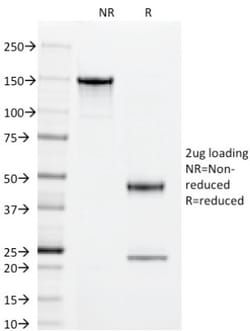
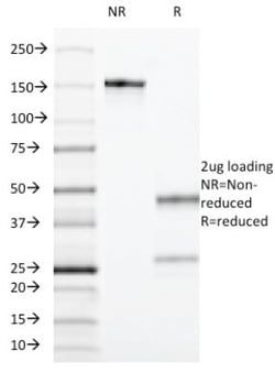
Recognizes a single chain glycoprotein of 70kDa, identified as CD55 (also known as decay accelerating factor, DAF). CD55/DAF is widely expressed on cells throughout the body including leukocytes, erythrocytes, epithelium, endothelium, and fibroblasts. It is a Glycosyl phosphatidylinositol anchored (GPI-anchored) member of the membrane bound complement regulatory proteins that inhibit autologous complement cascade activation. It prevents the amplification steps of the complement cascade by interfering with the assembly of the C3-convertases, C4b2a and C3bBb, and the C5-convertase, C4b2a3b and C3bBb3b. CD55 also serves as receptor for CD97 and for echovirus and Coxsackie B virus. The MAb 143-30 can be used as marker for paroxysmal nocturnal hemoglobinuria (PNH).
Content And Storage
Store at 4C.
Isotype
IgG1 κ
Applications
Flow Cytometry, Immunohistochemistry (Frozen), SDS-Page, Immunofluorescence
Clone
143-30
Conjugate
Unconjugated
Formulation
10mM PBS and 0.05% BSA with 0.05% Sodium Azide
Gene Alias
CD55 antigen, CD55 molecule, decay accelerating factor for complement (Cromer blood group), CRdecay accelerating factor for complement (CD55, Cromer blood group system), CROMDAFcomplement decay-accelerating factor, decay accelerating factor for complement, TC
Host Species
Mouse
Molecular Weight of Antigen
70 kDa
Quantity
0.1 mg
Research Discipline
Immunology
Gene ID (Entrez)
1604
Target Species
Human
Form
Purified
Related Products


Description
- Ensure accurate, reproducible results in Flow Cytometry, Immunohistochemistry (Frozen), Immunofluorescence CD55/DAF Monoclonal specifically detects CD55/DAF in Human samples
- It is validated for Flow Cytometry, Immunocytochemistry/Immunofluorescence, Flow (Cell Surface).
Compare Similar Items
Show Difference
Antigen: CD55/DAF
Classification: Monoclonal
Concentration: 0.2 mg/mL
Dilution: Flow Cytometry 0.5 - 1 ug/million cells in 0.1 ml, Immunohistochemistry-Frozen 0.5 - 1.0 ug/ml, SDS-Page, Immunofluorescence 0.5 - 1.0 ug/ml
Gene Accession No.: P08174, P08174
Gene Symbols: CD55
Immunogen: PHA stimulated human PBL
Purification Method: Protein A or G purified
Regulatory Status: RUO
Primary or Secondary: Primary
Test Specificity: Recognizes a single chain glycoprotein of 70kDa, identified as CD55 (also known as decay accelerating factor, DAF). CD55/DAF is widely expressed on cells throughout the body including leukocytes, erythrocytes, epithelium, endothelium, and fibroblasts. It is a Glycosyl phosphatidylinositol anchored (GPI-anchored) member of the membrane bound complement regulatory proteins that inhibit autologous complement cascade activation. It prevents the amplification steps of the complement cascade by interfering with the assembly of the C3-convertases, C4b2a and C3bBb, and the C5-convertase, C4b2a3b and C3bBb3b. CD55 also serves as receptor for CD97 and for echovirus and Coxsackie B virus. The MAb 143-30 can be used as marker for paroxysmal nocturnal hemoglobinuria (PNH).
Content And Storage: Store at 4C.
Isotype: IgG1 κ
Applications: Flow Cytometry, Immunohistochemistry (Frozen), SDS-Page, Immunofluorescence
Clone: 143-30
Conjugate: Unconjugated
Formulation: 10mM PBS and 0.05% BSA with 0.05% Sodium Azide
Gene Alias: CD55 antigen, CD55 molecule, decay accelerating factor for complement (Cromer blood group), CRdecay accelerating factor for complement (CD55, Cromer blood group system), CROMDAFcomplement decay-accelerating factor, decay accelerating factor for complement, TC
Host Species: Mouse
Molecular Weight of Antigen: 70 kDa
Quantity: 0.1 mg
Research Discipline: Immunology
Gene ID (Entrez): 1604
Target Species: Human
Form: Purified
Antigen: CD55/DAF
Classification: Monoclonal
Concentration: 0.2 mg/mL
Dilution: Flow Cytometry 0.5 - 1 ug/million cells in 0.1 ml, Immunohistochemistry-Frozen 0.5 - 1.0 ug/ml, SDS-Page, Immunofluorescence 0.5 - 1.0 ug/ml
Gene Accession No.: P08174, P08174
Gene Symbols: CD55
Immunogen: PHA stimulated human PBL
Purification Method: Protein A or G purified
Regulatory Status: RUO
Primary or Secondary: Primary
Test Specificity: Recognizes a single chain glycoprotein of 70kDa, identified as CD55 (also known as decay accelerating factor, DAF). CD55/DAF is widely expressed on cells throughout the body including leukocytes, erythrocytes, epithelium, endothelium, and fibroblasts. It is a Glycosyl phosphatidylinositol anchored (GPI-anchored) member of the membrane bound complement regulatory proteins that inhibit autologous complement cascade activation. It prevents the amplification steps of the complement cascade by interfering with the assembly of the C3-convertases, C4b2a and C3bBb, and the C5-convertase, C4b2a3b and C3bBb3b. CD55 also serves as receptor for CD97 and for echovirus and Coxsackie B virus. The MAb 143-30 can be used as marker for paroxysmal nocturnal hemoglobinuria (PNH).
Content And Storage: Store at 4C.
Isotype: IgG1 κ
Applications: Flow Cytometry, Immunohistochemistry (Frozen), SDS-Page, Immunofluorescence
Clone: 143-30
Conjugate: Unconjugated
Formulation: 10mM PBS and 0.05% BSA with 0.05% Sodium Azide
Gene Alias: CD55 antigen, CD55 molecule, decay accelerating factor for complement (Cromer blood group), CRdecay accelerating factor for complement (CD55, Cromer blood group system), CROMDAFcomplement decay-accelerating factor, decay accelerating factor for complement, TC
Host Species: Mouse
Molecular Weight of Antigen: 70 kDa
Quantity: 0.2 mg
Research Discipline: Immunology
Gene ID (Entrez): 1604
Target Species: Human
Form: Purified
Antigen: NGF R/TNFRSF16/p75NTR
Classification: Monoclonal
Concentration: 0.2mg/mL
Dilution: Flow Cytometry 0.5 - 1 ug/million cells in 0.1 ml, Immunocytochemistry/Immunofluorescence 1:10-1:500, Immunohistochemistry-Paraffin 0.5 - 1.0 ug/ml, Immunofluorescence 1 - 2 ug/ml
Gene Accession No.: P08138
Gene Symbols: NGFR
Immunogen: NGFR from A875 melanoma cells
Purification Method: Protein A or G purified
Regulatory Status: RUO
Primary or Secondary: Primary
Test Specificity: It recognizes a glycoprotein of 75kDa, identified as low affinity Nerve Growth Factor (NGF) Receptor (p75NGFR) or Neurotrophin Receptor (p75NTR). Its epitope spans in aa 1-160 of extracellular domain of NGFR/NTR. NGF-receptor contains an extracellular domain containing four 40-amino acid repeats with 6 cysteine residues at conserved positions followed by a serine/threonine-rich region, a single transmembrane domain, and a 155-amino acid cytoplasmic domain. The cysteine-rich region contains the nerve growth factor binding domain. NGF is important for the development, differentiation, and survival of variety of neuronal and non-neuronal cells. Its action is mediated by binding two distinct receptors, the high affinity p140 and low affinity p75.
Content And Storage: Store at 4C.
Isotype: IgG1 κ
Applications: Flow Cytometry, Immunocytochemistry, Immunofluorescence, Immunohistochemistry (Paraffin)
Clone: NGFR5
Conjugate: Unconjugated
Formulation: 10mM PBS and 0.05% BSA with 0.05% Sodium Azide
Gene Alias: CD271, CD271 antigen, Gp80-LNGFR, member 16, nerve growth factor receptor, NGF receptor, p75 ICD, TNFRSF16nerve growth factor receptor (TNFR superfamily, member 16), tumor necrosis factor receptor superfamily member 16
Host Species: Mouse
Molecular Weight of Antigen: 75 kDa
Quantity: 0.02 mg
Research Discipline: Apoptosis, Neuronal Cell Markers, Neuronal Stem Cell Markers, Neuroscience, Stem Cell Markers
Gene ID (Entrez): 4804
Target Species: Human, Baboon, Feline, Ferret, Monkey, Rabbit, Mouse (Negative), Rat (Negative)
Form: Purified