NF-L Antibody (NFL/736), Novus Biologicals™
Manufacturer: Fischer Scientific
The price for this product is unavailable. Please request a quote
Antigen
NF-L
Classification
Monoclonal
Concentration
0.2mg/mL
Dilution
Immunohistochemistry-Paraffin 0.25 - 0.5 ug/ml, Immunofluorescence 1 - 2 ug/ml
Gene Accession No.
P07196
Gene Symbols
NEFL
Immunogen
Recombinant human NEFL protein
Purification Method
Protein A or G purified
Regulatory Status
RUO
Primary or Secondary
Primary
Test Specificity
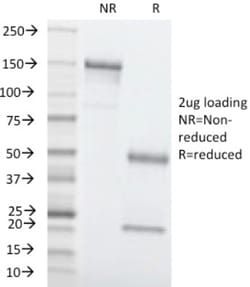
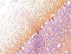
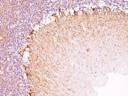
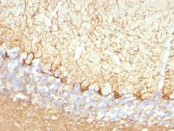
This MAb reacts with a 68kDa protein, identified as light sub-unit of neurofilaments (NF-L). Neurofilaments make up the main structural elements of axons and dendrites and are found in neurons, peripheral nerves, and sympathetic ganglion cells. Neurofilaments consist of three major subunits with molecular weights of 68kDa (NF-L), 160kDa (NF-M) and 200kDa (NF-H). Anti-neurofilament stains a number of neural, neuroendocrine, and endocrine tumors. Neuromas, ganglioneuromas, gangliogliomas, ganglioneuroblastomas, and neuroblastomas stain positively for anti-neurofilament. Neurofilaments are also present in paragangliomas as well as adrenal and extra-adrenal pheochromocytomas. Carcinoids, neuroendocrine carcinomas of the skin, and oat cell carcinomas of the lung also express neurofilament.
Content And Storage
Store at 4C.
Isotype
IgG1
Applications
Immunohistochemistry (Paraffin), Immunofluorescence
Clone
NFL/736
Conjugate
Unconjugated
Formulation
10mM PBS and 0.05% BSA with 0.05% Sodium Azide
Gene Alias
68 kDa neurofilament protein, neurofilament protein, light chain, neurofilament subunit NF-L, Neurofilament triplet L protein, neurofilament, light polypeptide, neurofilament-light, NF68FLJ53642, NF-L, NFLlight polypeptide 68kDa
Host Species
Mouse
Molecular Weight of Antigen
68 kDa
Quantity
0.2 mg
Research Discipline
Autophagy, Cellular Markers, Cytoskeleton Markers, Neurodegeneration, Neurofilaments, Neuronal Cell Markers, Neuroscience
Gene ID (Entrez)
4747
Target Species
Human, Rat, Porcine, Chicken, Primate
Form
Purified
Related Products

Description
- Ensure accurate, reproducible results in Immunohistochemistry (Paraffin), Immunofluorescence NF-L Monoclonal specifically detects NF-L in Human, Rat, Porcine, Bovine, Chicken samples
- It is validated for Flow Cytometry, Immunohistochemistry, Immunocytochemistry/Immunofluorescence, Immunohistochemistry-Paraffin, Immunofluorescence.
Compare Similar Items
Show Difference
Antigen: NF-L
Classification: Monoclonal
Concentration: 0.2mg/mL
Dilution: Immunohistochemistry-Paraffin 0.25 - 0.5 ug/ml, Immunofluorescence 1 - 2 ug/ml
Gene Accession No.: P07196
Gene Symbols: NEFL
Immunogen: Recombinant human NEFL protein
Purification Method: Protein A or G purified
Regulatory Status: RUO
Primary or Secondary: Primary
Test Specificity: This MAb reacts with a 68kDa protein, identified as light sub-unit of neurofilaments (NF-L). Neurofilaments make up the main structural elements of axons and dendrites and are found in neurons, peripheral nerves, and sympathetic ganglion cells. Neurofilaments consist of three major subunits with molecular weights of 68kDa (NF-L), 160kDa (NF-M) and 200kDa (NF-H). Anti-neurofilament stains a number of neural, neuroendocrine, and endocrine tumors. Neuromas, ganglioneuromas, gangliogliomas, ganglioneuroblastomas, and neuroblastomas stain positively for anti-neurofilament. Neurofilaments are also present in paragangliomas as well as adrenal and extra-adrenal pheochromocytomas. Carcinoids, neuroendocrine carcinomas of the skin, and oat cell carcinomas of the lung also express neurofilament.
Content And Storage: Store at 4C.
Isotype: IgG1
Applications: Immunohistochemistry (Paraffin), Immunofluorescence
Clone: NFL/736
Conjugate: Unconjugated
Formulation: 10mM PBS and 0.05% BSA with 0.05% Sodium Azide
Gene Alias: 68 kDa neurofilament protein, neurofilament protein, light chain, neurofilament subunit NF-L, Neurofilament triplet L protein, neurofilament, light polypeptide, neurofilament-light, NF68FLJ53642, NF-L, NFLlight polypeptide 68kDa
Host Species: Mouse
Molecular Weight of Antigen: 68 kDa
Quantity: 0.2 mg
Research Discipline: Autophagy, Cellular Markers, Cytoskeleton Markers, Neurodegeneration, Neurofilaments, Neuronal Cell Markers, Neuroscience
Gene ID (Entrez): 4747
Target Species: Human, Rat, Porcine, Chicken, Primate
Form: Purified
Antigen: CEACAM5/CD66e
Classification: Monoclonal
Concentration: 0.2mg/mL
Dilution: Flow Cytometry 0.5 - 1 ug/million cells in 0.1 ml, Immunohistochemistry-Paraffin 1 - 2 ug/ml, SDS-Page, Immunofluorescence 1 - 2 ug/ml
Gene Accession No.: P06731
Gene Symbols: CEACAM5
Immunogen: Purified human CEA protein
Purification Method: Protein A or G purified
Regulatory Status: RUO
Primary or Secondary: Primary
Test Specificity: This antibody recognizes proteins of 80-200kDa, identified as different members of CEA family. CEA is synthesized during development in the fetal gut and is re-expressed in increased amounts in intestinal carcinomas and several other tumors. This MAb reacts with nonspecific cross-reacting antigen (NCA). It shows no reaction with a variety of normal tissues and is suitable for staining of formalin/paraffin tissues. CEA is not found in benign glands, stroma, or malignant prostatic cells. Antibody to CEA is useful in detecting early foci of gastric carcinoma and in distinguishing pulmonary adenocarcinomas (60-70% are CEA+) from pleural mesotheliomas (rarely or weakly CEA+). Anti-CEA positivity is seen in adenocarcinomas from the lung, colon, stomach, esophagus, pancreas, gallbadder, urachus, salivary gland, ovary, and endocervix.
Content And Storage: Store at 4C.
Isotype: IgG2a κ
Applications: Flow Cytometry, Immunohistochemistry (Paraffin), SDS-Page, Immunofluorescence
Clone: C66/1260
Conjugate: Unconjugated
Formulation: 10mM PBS and 0.05% BSA with 0.05% Sodium Azide
Gene Alias: Carcinoembryonic antigen, carcinoembryonic antigen-related cell adhesion molecule 5, CD66e antigen, CEACD66e, DKFZp781M2392, Meconium antigen 100
Host Species: Mouse
Molecular Weight of Antigen: __
Quantity: 0.02 mg
Research Discipline: Cancer, Cellular Markers, Immunology
Gene ID (Entrez): 1048
Target Species: Human
Form: Purified
Antigen: CEACAM5/CD66e
Classification: Monoclonal
Concentration: 0.2mg/mL
Dilution: Flow Cytometry 0.5 - 1 ug/million cells in 0.1 ml, Immunohistochemistry-Paraffin 1 - 2 ug/ml, SDS-Page, Immunofluorescence 1 - 2 ug/ml
Gene Accession No.: P06731
Gene Symbols: CEACAM5
Immunogen: Purified human CEA protein
Purification Method: Protein A or G purified
Regulatory Status: RUO
Primary or Secondary: Primary
Test Specificity: This antibody recognizes proteins of 80-200kDa, identified as different members of CEA family. CEA is synthesized during development in the fetal gut and is re-expressed in increased amounts in intestinal carcinomas and several other tumors. This MAb reacts with nonspecific cross-reacting antigen (NCA). It shows no reaction with a variety of normal tissues and is suitable for staining of formalin/paraffin tissues. CEA is not found in benign glands, stroma, or malignant prostatic cells. Antibody to CEA is useful in detecting early foci of gastric carcinoma and in distinguishing pulmonary adenocarcinomas (60-70% are CEA+) from pleural mesotheliomas (rarely or weakly CEA+). Anti-CEA positivity is seen in adenocarcinomas from the lung, colon, stomach, esophagus, pancreas, gallbadder, urachus, salivary gland, ovary, and endocervix.
Content And Storage: Store at 4C.
Isotype: IgG2a κ
Applications: Flow Cytometry, Immunohistochemistry (Paraffin), SDS-Page, Immunofluorescence
Clone: C66/1260
Conjugate: Unconjugated
Formulation: 10mM PBS and 0.05% BSA with 0.05% Sodium Azide
Gene Alias: Carcinoembryonic antigen, carcinoembryonic antigen-related cell adhesion molecule 5, CD66e antigen, CEACD66e, DKFZp781M2392, Meconium antigen 100
Host Species: Mouse
Molecular Weight of Antigen: __
Quantity: 0.1 mg
Research Discipline: Cancer, Cellular Markers, Immunology
Gene ID (Entrez): 1048
Target Species: Human
Form: Purified