CD2 Antibody (HuLy-m1), Novus Biologicals™
Manufacturer: Fischer Scientific
The price for this product is unavailable. Please request a quote
Antigen
CD2
Classification
Monoclonal
Concentration
0.2 mg/mL
Dilution
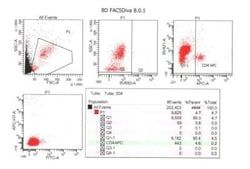
Flow Cytometry 0.5 - 1 ug/million cells in 0.1 ml, Immunofluorescence 0.5 - 1.0 ug/ml
Gene Accession No.
P06729
Gene Symbols
CD2
Immunogen
Human thymocytes
Purification Method
Protein A or G purified
Regulatory Status
RUO
Primary or Secondary
Primary
Test Specificity
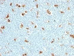
CD2 interacts through its amino-terminal domain with the extracellular domain of CD58 (also designated CD2 ligand) to mediate cell adhesion. CD2/CD58 binding can enhance antigen-specific T cell activation. CD2 is a transmembrane glycoprotein that is expressed on peripheral blood T lymphocytes, NK cells and thymocytes. CD58 is a heavily glycosylated protein with a broad tissue distribution in hematopoietic and other cells, including endothelium. Interaction between CD2 and its counter receptor LFA3 (CD58) on opposing cells optimizes immune system recognition, thereby facilitating communication between helper T lymphocytes and antigen-presenting cells, as well as between cytolytic effectors and target cells.
Content And Storage
Store at 4C.
Isotype
IgG2b κ
Applications
Flow Cytometry, Immunofluorescence
Clone
HuLy-m1
Conjugate
Unconjugated
Formulation
10mM PBS and 0.05% BSA with 0.05% Sodium Azide
Gene Alias
CD2 antigen, CD2 antigen (p50), sheep red blood cell receptor, CD2 molecule, Erythrocyte receptor, FLJ46032, LFA-2, LFA-3 receptor, lymphocyte-function antigen-2, Rosette receptor, SRBC, T11, T-cell surface antigen CD2, T-cell surface antigen T11/Leu-5
Host Species
Mouse
Molecular Weight of Antigen
50 kDa
Quantity
0.2 mg
Research Discipline
Adaptive Immunity, Apoptosis, Immunology
Gene ID (Entrez)
914
Target Species
Human, Feline
Form
Purified
Related Products


Description
- Ensure accurate, reproducible results in Flow Cytometry, Immunofluorescence CD2 Monoclonal specifically detects CD2 in Human, Feline samples
- It is validated for Flow Cytometry, Immunocytochemistry/Immunofluorescence, Functional, Immunofluorescence.
Compare Similar Items
Show Difference
Antigen: CD2
Classification: Monoclonal
Concentration: 0.2 mg/mL
Dilution: Flow Cytometry 0.5 - 1 ug/million cells in 0.1 ml, Immunofluorescence 0.5 - 1.0 ug/ml
Gene Accession No.: P06729
Gene Symbols: CD2
Immunogen: Human thymocytes
Purification Method: Protein A or G purified
Regulatory Status: RUO
Primary or Secondary: Primary
Test Specificity: CD2 interacts through its amino-terminal domain with the extracellular domain of CD58 (also designated CD2 ligand) to mediate cell adhesion. CD2/CD58 binding can enhance antigen-specific T cell activation. CD2 is a transmembrane glycoprotein that is expressed on peripheral blood T lymphocytes, NK cells and thymocytes. CD58 is a heavily glycosylated protein with a broad tissue distribution in hematopoietic and other cells, including endothelium. Interaction between CD2 and its counter receptor LFA3 (CD58) on opposing cells optimizes immune system recognition, thereby facilitating communication between helper T lymphocytes and antigen-presenting cells, as well as between cytolytic effectors and target cells.
Content And Storage: Store at 4C.
Isotype: IgG2b κ
Applications: Flow Cytometry, Immunofluorescence
Clone: HuLy-m1
Conjugate: Unconjugated
Formulation: 10mM PBS and 0.05% BSA with 0.05% Sodium Azide
Gene Alias: CD2 antigen, CD2 antigen (p50), sheep red blood cell receptor, CD2 molecule, Erythrocyte receptor, FLJ46032, LFA-2, LFA-3 receptor, lymphocyte-function antigen-2, Rosette receptor, SRBC, T11, T-cell surface antigen CD2, T-cell surface antigen T11/Leu-5
Host Species: Mouse
Molecular Weight of Antigen: 50 kDa
Quantity: 0.2 mg
Research Discipline: Adaptive Immunity, Apoptosis, Immunology
Gene ID (Entrez): 914
Target Species: Human, Feline
Form: Purified
Antigen: S100A9
Classification: Monoclonal
Concentration: 0.2 mg/mL
Dilution: Flow Cytometry 0.5 - 1 ug/million cells in 0.1 ml, Immunohistochemistry-Paraffin 0.5 - 1 ug/ml, Immunohistochemistry-Frozen (Negative), Immunofluorescence 0.5 - 1.0 ug/ml
Gene Accession No.: __
Gene Symbols: S100A9
Immunogen: Recombinant human S100A9 protein
Purification Method: Protein A or G purified
Regulatory Status: RUO
Primary or Secondary: Primary
Test Specificity: Recognizes a 14kDa protein, identified as S100A9 (also known as Calgranulin B or MRP-14); expressed by granulocytes, monocytes and by tissue macrophages.The protein encoded by this gene is a member of the S100 family of proteins containing 2 EF-hand calcium-binding motifs. S100 proteins are localized in the cytoplasm and/or nucleus of a wide range of cells, and involved in the regulation of a number of cellular processes such as cell cycle progression and differentiation. Altered expression of this protein is associated with the disease cystic fibrosis. This MAb reacts with neutrophils, monocytes and macrophages, and has been shown as an important marker for identifying macrophages in tissue sections. Among cells that are now recognized as macrophages are histiocytes, Kupffer cells, osteoclasts, microglial cells, synovial type A cells, interdigitating cells, and Langerhans cells (in normal tissues) and epithelioid cells and Langerhans-type and foreign-body-type multinucleated giant cel
Content And Storage: Store at 4C.
Isotype: IgM κ
Applications: Flow Cytometry, Immunohistochemistry (Paraffin), Immunohistochemistry (Frozen), Immunofluorescence
Clone: S100A9/1011
Conjugate: Unconjugated
Formulation: 10mM PBS with 0.05% Sodium Azide
Gene Alias: CAGBMigration inhibitory factor-related protein 14, calgranulin B, calgranulin-B, Calprotectin L1H subunit, CFAGMRP-14,60B8AG, CGLB, L1AG, LIAG, MAC387, MIF, MRP14Leukocyte L1 complex heavy chain, NIF, P14, protein S100-A9, S100 calcium binding protein A9, S100 calcium binding protein A9 (calgranulin B), S100 calcium-binding protein A9, S100 calcium-binding protein A9 (calgranulin B)
Host Species: Mouse
Molecular Weight of Antigen: 14 kDa
Quantity: 0.02 mg
Research Discipline: Cancer, Immunology, Innate Immunity, Signal Transduction, Tyrosine Kinases
Gene ID (Entrez): 6280
Target Species: Human
Form: Purified
Antigen: S100A9
Classification: Monoclonal
Concentration: 0.2 mg/mL
Dilution: Flow Cytometry 0.5 - 1 ug/million cells in 0.1 ml, Immunohistochemistry-Paraffin 0.5 - 1 ug/ml, Immunohistochemistry-Frozen (Negative), Immunofluorescence 0.5 - 1.0 ug/ml
Gene Accession No.: __
Gene Symbols: S100A9
Immunogen: Recombinant human S100A9 protein
Purification Method: Protein A or G purified
Regulatory Status: RUO
Primary or Secondary: Primary
Test Specificity: Recognizes a 14kDa protein, identified as S100A9 (also known as Calgranulin B or MRP-14); expressed by granulocytes, monocytes and by tissue macrophages.The protein encoded by this gene is a member of the S100 family of proteins containing 2 EF-hand calcium-binding motifs. S100 proteins are localized in the cytoplasm and/or nucleus of a wide range of cells, and involved in the regulation of a number of cellular processes such as cell cycle progression and differentiation. Altered expression of this protein is associated with the disease cystic fibrosis. This MAb reacts with neutrophils, monocytes and macrophages, and has been shown as an important marker for identifying macrophages in tissue sections. Among cells that are now recognized as macrophages are histiocytes, Kupffer cells, osteoclasts, microglial cells, synovial type A cells, interdigitating cells, and Langerhans cells (in normal tissues) and epithelioid cells and Langerhans-type and foreign-body-type multinucleated giant cel
Content And Storage: Store at 4C.
Isotype: IgM κ
Applications: Flow Cytometry, Immunohistochemistry (Paraffin), Immunohistochemistry (Frozen), Immunofluorescence
Clone: S100A9/1011
Conjugate: Unconjugated
Formulation: 10mM PBS with 0.05% Sodium Azide
Gene Alias: CAGBMigration inhibitory factor-related protein 14, calgranulin B, calgranulin-B, Calprotectin L1H subunit, CFAGMRP-14,60B8AG, CGLB, L1AG, LIAG, MAC387, MIF, MRP14Leukocyte L1 complex heavy chain, NIF, P14, protein S100-A9, S100 calcium binding protein A9, S100 calcium binding protein A9 (calgranulin B), S100 calcium-binding protein A9, S100 calcium-binding protein A9 (calgranulin B)
Host Species: Mouse
Molecular Weight of Antigen: 14 kDa
Quantity: 0.1 mg
Research Discipline: Cancer, Immunology, Innate Immunity, Signal Transduction, Tyrosine Kinases
Gene ID (Entrez): 6280
Target Species: Human
Form: Purified