Von Willebrand Factor Antibody (IIIE2.34), Novus Biologicals™
Manufacturer: Fischer Scientific
Select a Size
| Pack Size | SKU | Availability | Price |
|---|---|---|---|
| Each of 1 | NBP24499501-Each-of-1 | In Stock | ₹ 46,636.00 |
NBP24499501 - Each of 1
In Stock
Quantity
1
Base Price: ₹ 46,636.00
GST (18%): ₹ 8,394.48
Total Price: ₹ 55,030.48
Antigen
Von Willebrand Factor
Classification
Monoclonal
Concentration
0.2 mg/mL
Dilution
Western Blot 0.5 - 1.0 ug/ml, Flow Cytometry 0.5 - 1 ug/million cells in 0.1 ml, Immunoprecipitation 0.5 - 1 ug/500 ug Protein Lysate, Immunohistochemistry-Paraffin 0.5 - 1.0 ug/ml, Immunofluorescence 0.5 - 1.0 ug/ml
Gene Accession No.
P04275
Gene Symbols
VWF
Immunogen
Recombinant human vWF fragment spanning aa 845-949
Purification Method
Protein A or G purified
Regulatory Status
RUO
Primary or Secondary
Primary
Test Specificity
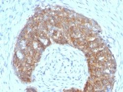
Von Willebrand Factor (vWF) is a multimeric glycoprotein that is found in endothelial cells, plasma and platelets. It acts as a carrier protein for Factor VIII and promotes platelet adhesion and aggregation. vWF undergoes a variety of posttranslational modifications that influence the affinity and availability for Factor VIII, including cleavage of the propeptide and formation of N-terminal disulfide bonds. This antibody helps to establish the endothelial nature of some lesions of disputed histogenesis, e.g. Kaposi's sarcoma and cardiac myxoma. It is widely used for differentiating vascular lesions from those of other tissue differentiation within a panel of other vascular markers although not all tumors of endothelial differentiation contain this antigen.
Content And Storage
Store at 4C.
Isotype
IgG1 κ
Applications
Western Blot, Flow Cytometry, Immunoprecipitation, Immunohistochemistry (Paraffin), Immunofluorescence
Clone
IIIE2.34
Conjugate
Unconjugated
Formulation
10mM PBS and 0.05% BSA with 0.05% Sodium Azide
Gene Alias
coagulation factor VIII VWF, F8, F8VWF, von Willebrand factor, VWD, vWF
Host Species
Mouse
Molecular Weight of Antigen
250 kDa
Quantity
0.1 mg
Research Discipline
Cancer
Gene ID (Entrez)
7450
Target Species
Human
Form
Purified
Related Products


Description
- Ensure accurate, reproducible results in Western Blot, Flow Cytometry, Immunohistochemistry (Paraffin), Immunoprecipitation, Immunofluorescence Von Willebrand Factor Monoclonal specifically detects Von Willebrand Factor in Human samples
- It is validated for Western Blot, Flow Cytometry, Immunohistochemistry, Immunocytochemistry/Immunofluorescence, Immunoprecipitation, Immunohistochemistry-Paraffin, Immunofluorescence.
Compare Similar Items
Show Difference
Antigen: Von Willebrand Factor
Classification: Monoclonal
Concentration: 0.2 mg/mL
Dilution: Western Blot 0.5 - 1.0 ug/ml, Flow Cytometry 0.5 - 1 ug/million cells in 0.1 ml, Immunoprecipitation 0.5 - 1 ug/500 ug Protein Lysate, Immunohistochemistry-Paraffin 0.5 - 1.0 ug/ml, Immunofluorescence 0.5 - 1.0 ug/ml
Gene Accession No.: P04275
Gene Symbols: VWF
Immunogen: Recombinant human vWF fragment spanning aa 845-949
Purification Method: Protein A or G purified
Regulatory Status: RUO
Primary or Secondary: Primary
Test Specificity: Von Willebrand Factor (vWF) is a multimeric glycoprotein that is found in endothelial cells, plasma and platelets. It acts as a carrier protein for Factor VIII and promotes platelet adhesion and aggregation. vWF undergoes a variety of posttranslational modifications that influence the affinity and availability for Factor VIII, including cleavage of the propeptide and formation of N-terminal disulfide bonds. This antibody helps to establish the endothelial nature of some lesions of disputed histogenesis, e.g. Kaposi's sarcoma and cardiac myxoma. It is widely used for differentiating vascular lesions from those of other tissue differentiation within a panel of other vascular markers although not all tumors of endothelial differentiation contain this antigen.
Content And Storage: Store at 4C.
Isotype: IgG1 κ
Applications: Western Blot, Flow Cytometry, Immunoprecipitation, Immunohistochemistry (Paraffin), Immunofluorescence
Clone: IIIE2.34
Conjugate: Unconjugated
Formulation: 10mM PBS and 0.05% BSA with 0.05% Sodium Azide
Gene Alias: coagulation factor VIII VWF, F8, F8VWF, von Willebrand factor, VWD, vWF
Host Species: Mouse
Molecular Weight of Antigen: 250 kDa
Quantity: 0.1 mg
Research Discipline: Cancer
Gene ID (Entrez): 7450
Target Species: Human
Form: Purified
Antigen: TFF1/pS2
Classification: Monoclonal
Concentration: 0.2mg/mL
Dilution: Flow Cytometry 0.5 - 1 ug/million cells in 0.1 ml, Immunohistochemistry-Paraffin 0.5 - 1.0 ug/ml, Immunofluorescence 0.5 - 1.0 ug/ml
Gene Accession No.: P04155
Gene Symbols: TFF1
Immunogen: Synthetic peptide of 28 amino acid residues corresponding to CFDDTVRGVPWCFYPNTIDVPPEEECEF (aa57-84) from the C-terminus of human pS2.
Purification Method: Protein A or G purified
Regulatory Status: RUO
Primary or Secondary: Primary
Test Specificity: It recognizes a polypeptide of 6.5kDa, identified as pS2 estrogen-regulated protein. Its epitope is localized between aa57-84 of human pS2 protein. pS2 is a trefoil peptide. Trefoil peptides are protease resistant molecules secreted throughout the gut that play a role in mucosal healing. These peptides contain three intra-chain disulfide bonds, forming the trefoil motif, or P-domain. pS2 is known to form dimers and this dimerization is thought to play a role in its protective and healing properties. About 60% of breast carcinomas are positive for pS2. Staining is cytoplasmic, often with localization to the Golgi apparatus. pS2 is shown to be localized in normal stomach mucosa, gastric fluid, goblet cells in the colon and small intestine, and in ulcerations of the gastrointestinal tract. Several studies have shown that pS2 is primarily expressed in estrogen receptor-positive breast tumors and it may define a subset of estrogen-dependent tumors that displays an increased likelihood of re
Content And Storage: Store at 4C.
Isotype: IgG1 κ
Applications: Flow Cytometry, Immunohistochemistry (Paraffin), Immunofluorescence
Clone: SPM313
Conjugate: Unconjugated
Formulation: 10mM PBS and 0.05% BSA with 0.05% Sodium Azide
Gene Alias: BCEIbreast cancer, estrogen-inducible sequence expressed in, Breast cancer estrogen-inducible protein, breast cancer estrogen-inducible sequence, D21S21, gastrointestinal trefoil protein pS2, hP1.A, HPS2, pNR-2, Polypeptide P1.A, Protein pS2, pS2, trefoil factor 1, trefoil factor, BCE1, human pS2 induced by estrogen from human breast cancercell line M10HP1.A
Host Species: Mouse
Molecular Weight of Antigen: 6.5 kDa
Quantity: 0.02 mg
Research Discipline: Breast Cancer, Cancer
Gene ID (Entrez): 7031
Target Species: Human, Cynomolgus Monkey
Form: Purified
Antigen: TFF1/pS2
Classification: Monoclonal
Concentration: 0.2mg/mL
Dilution: Flow Cytometry 0.5 - 1 ug/million cells in 0.1 ml, Immunohistochemistry-Paraffin 0.5 - 1.0 ug/ml, Immunofluorescence 0.5 - 1.0 ug/ml
Gene Accession No.: P04155
Gene Symbols: TFF1
Immunogen: Synthetic peptide of 28 amino acid residues corresponding to CFDDTVRGVPWCFYPNTIDVPPEEECEF (aa57-84) from the C-terminus of human pS2.
Purification Method: Protein A or G purified
Regulatory Status: RUO
Primary or Secondary: Primary
Test Specificity: It recognizes a polypeptide of 6.5kDa, identified as pS2 estrogen-regulated protein. Its epitope is localized between aa57-84 of human pS2 protein. pS2 is a trefoil peptide. Trefoil peptides are protease resistant molecules secreted throughout the gut that play a role in mucosal healing. These peptides contain three intra-chain disulfide bonds, forming the trefoil motif, or P-domain. pS2 is known to form dimers and this dimerization is thought to play a role in its protective and healing properties. About 60% of breast carcinomas are positive for pS2. Staining is cytoplasmic, often with localization to the Golgi apparatus. pS2 is shown to be localized in normal stomach mucosa, gastric fluid, goblet cells in the colon and small intestine, and in ulcerations of the gastrointestinal tract. Several studies have shown that pS2 is primarily expressed in estrogen receptor-positive breast tumors and it may define a subset of estrogen-dependent tumors that displays an increased likelihood of re
Content And Storage: Store at 4C.
Isotype: IgG1 κ
Applications: Flow Cytometry, Immunohistochemistry (Paraffin), Immunofluorescence
Clone: SPM313
Conjugate: Unconjugated
Formulation: 10mM PBS and 0.05% BSA with 0.05% Sodium Azide
Gene Alias: BCEIbreast cancer, estrogen-inducible sequence expressed in, Breast cancer estrogen-inducible protein, breast cancer estrogen-inducible sequence, D21S21, gastrointestinal trefoil protein pS2, hP1.A, HPS2, pNR-2, Polypeptide P1.A, Protein pS2, pS2, trefoil factor 1, trefoil factor, BCE1, human pS2 induced by estrogen from human breast cancercell line M10HP1.A
Host Species: Mouse
Molecular Weight of Antigen: 6.5 kDa
Quantity: 0.1 mg
Research Discipline: Breast Cancer, Cancer
Gene ID (Entrez): 7031
Target Species: Human, Cynomolgus Monkey
Form: Purified