Cytokeratin 14 Antibody (KRT14/532), Novus Biologicals™
Manufacturer: Fischer Scientific
Select a Size
| Pack Size | SKU | Availability | Price |
|---|---|---|---|
| Each of 1 | NBP24503201-Each-of-1 | In Stock | ₹ 46,636.00 |
NBP24503201 - Each of 1
In Stock
Quantity
1
Base Price: ₹ 46,636.00
GST (18%): ₹ 8,394.48
Total Price: ₹ 55,030.48
Antigen
Cytokeratin 14
Classification
Monoclonal
Concentration
0.2mg/mL
Dilution
Flow Cytometry 0.5 - 1 ug/million cells in 0.1 ml, Immunohistochemistry-Paraffin 0.5 - 1.0 ug/ml, Immunofluorescence 0.5 - 1.0 ug/ml
Gene Accession No.
P02533
Gene Symbols
KRT14
Immunogen
Recombinant full-length human KRT14 protein
Purification Method
Protein A or G purified
Regulatory Status
RUO
Primary or Secondary
Primary
Test Specificity
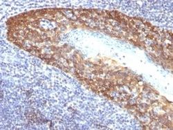
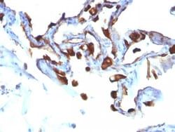
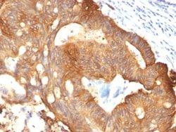
Cytokeratin 14 (CK14) belongs to the type I (or A or acidic) subfamily of low molecular weight keratins and exists in combination with keratin 5 (type II or B or basic). CK14 is found in basal cells of squamous epithelia, some glandular epithelia, myoepithelium, and mesothelial cells. Anti-CK14 is useful in differentiating squamous cell carcinomas from poorly differentiated epithelial tumors. Anti-CK14 is one of the specific basal markers for distinguishing between basal and non-basal subtypes of breast carcinomas. Anti-CK14 is also a good marker for differentiation of intraductal from invasive salivary duct carcinoma by the positive staining of basal cells surrounding the in-situ neoplasm as well as for differentiation of benign prostate from prostate carcinoma. Furthermore, this antibody has been useful in separating oncocytic tumors of the kidney from its renal mimics, and in identifying metaplastic carcinomas of the breast.
Content And Storage
Store at 4C.
Isotype
IgG3
Applications
Flow Cytometry, Immunohistochemistry (Paraffin), Immunofluorescence
Clone
KRT14/532
Conjugate
Unconjugated
Formulation
10mM PBS and 0.05% BSA with 0.05% Sodium Azide
Gene Alias
CK14, CK-14, cytokeratin 14, cytokeratin-14, EBS3, EBS4, K14, keratin 14, keratin 14 (epidermolysis bullosa simplex, Dowling-Meara, Koebner), keratin, type I cytoskeletal 14, Keratin-14, NFJ
Host Species
Mouse
Molecular Weight of Antigen
50 kDa
Quantity
0.1 mg
Research Discipline
Cytoskeleton Markers
Gene ID (Entrez)
3861
Target Species
Human, Mouse, Rat
Form
Purified
Related Products

Description
- Ensure accurate, reproducible results in Flow Cytometry, Immunohistochemistry (Paraffin), Immunofluorescence Cytokeratin 14 Monoclonal specifically detects Cytokeratin 14 in Human, Mouse, Rat samples
- It is validated for Flow Cytometry, Immunohistochemistry, Immunocytochemistry/Immunofluorescence, Immunohistochemistry-Paraffin, Immunofluorescence.
Compare Similar Items
Show Difference
Antigen: Cytokeratin 14
Classification: Monoclonal
Concentration: 0.2mg/mL
Dilution: Flow Cytometry 0.5 - 1 ug/million cells in 0.1 ml, Immunohistochemistry-Paraffin 0.5 - 1.0 ug/ml, Immunofluorescence 0.5 - 1.0 ug/ml
Gene Accession No.: P02533
Gene Symbols: KRT14
Immunogen: Recombinant full-length human KRT14 protein
Purification Method: Protein A or G purified
Regulatory Status: RUO
Primary or Secondary: Primary
Test Specificity: Cytokeratin 14 (CK14) belongs to the type I (or A or acidic) subfamily of low molecular weight keratins and exists in combination with keratin 5 (type II or B or basic). CK14 is found in basal cells of squamous epithelia, some glandular epithelia, myoepithelium, and mesothelial cells. Anti-CK14 is useful in differentiating squamous cell carcinomas from poorly differentiated epithelial tumors. Anti-CK14 is one of the specific basal markers for distinguishing between basal and non-basal subtypes of breast carcinomas. Anti-CK14 is also a good marker for differentiation of intraductal from invasive salivary duct carcinoma by the positive staining of basal cells surrounding the in-situ neoplasm as well as for differentiation of benign prostate from prostate carcinoma. Furthermore, this antibody has been useful in separating oncocytic tumors of the kidney from its renal mimics, and in identifying metaplastic carcinomas of the breast.
Content And Storage: Store at 4C.
Isotype: IgG3
Applications: Flow Cytometry, Immunohistochemistry (Paraffin), Immunofluorescence
Clone: KRT14/532
Conjugate: Unconjugated
Formulation: 10mM PBS and 0.05% BSA with 0.05% Sodium Azide
Gene Alias: CK14, CK-14, cytokeratin 14, cytokeratin-14, EBS3, EBS4, K14, keratin 14, keratin 14 (epidermolysis bullosa simplex, Dowling-Meara, Koebner), keratin, type I cytoskeletal 14, Keratin-14, NFJ
Host Species: Mouse
Molecular Weight of Antigen: 50 kDa
Quantity: 0.1 mg
Research Discipline: Cytoskeleton Markers
Gene ID (Entrez): 3861
Target Species: Human, Mouse, Rat
Form: Purified
Antigen: Cytokeratin 14
Classification: Monoclonal
Concentration: 0.2mg/mL
Dilution: Flow Cytometry 0.5 - 1 ug/million cells in 0.1 ml, Immunohistochemistry-Paraffin 0.5 - 1.0 ug/ml, Immunofluorescence 0.5 - 1.0 ug/ml
Gene Accession No.: P02533
Gene Symbols: KRT14
Immunogen: Recombinant full-length human KRT14 protein
Purification Method: Protein A or G purified
Regulatory Status: RUO
Primary or Secondary: Primary
Test Specificity: Cytokeratin 14 (CK14) belongs to the type I (or A or acidic) subfamily of low molecular weight keratins and exists in combination with keratin 5 (type II or B or basic). CK14 is found in basal cells of squamous epithelia, some glandular epithelia, myoepithelium, and mesothelial cells. Anti-CK14 is useful in differentiating squamous cell carcinomas from poorly differentiated epithelial tumors. Anti-CK14 is one of the specific basal markers for distinguishing between basal and non-basal subtypes of breast carcinomas. Anti-CK14 is also a good marker for differentiation of intraductal from invasive salivary duct carcinoma by the positive staining of basal cells surrounding the in-situ neoplasm as well as for differentiation of benign prostate from prostate carcinoma. Furthermore, this antibody has been useful in separating oncocytic tumors of the kidney from its renal mimics, and in identifying metaplastic carcinomas of the breast.
Content And Storage: Store at 4C.
Isotype: IgG3
Applications: Flow Cytometry, Immunohistochemistry (Paraffin), Immunofluorescence
Clone: KRT14/532
Conjugate: Unconjugated
Formulation: 10mM PBS and 0.05% BSA with 0.05% Sodium Azide
Gene Alias: CK14, CK-14, cytokeratin 14, cytokeratin-14, EBS3, EBS4, K14, keratin 14, keratin 14 (epidermolysis bullosa simplex, Dowling-Meara, Koebner), keratin, type I cytoskeletal 14, Keratin-14, NFJ
Host Species: Mouse
Molecular Weight of Antigen: 50 kDa
Quantity: 0.2 mg
Research Discipline: Cytoskeleton Markers
Gene ID (Entrez): 3861
Target Species: Human, Mouse, Rat
Form: Purified

Antigen: HLA DQ
Classification: Monoclonal
Concentration: 0.2 mg/mL
Dilution: Flow Cytometry 0.5 - 1 ug/million cells in 0.1 ml, Immunohistochemistry-Frozen 0.5-1ug/ml, Immunofluorescence 0.5 - 1.0 ug/ml
Gene Accession No.: P01908
Gene Symbols: HLA-DQ
Immunogen: T4-positive CTL clone HG-38
Purification Method: Protein A or G purified
Regulatory Status: RUO
Primary or Secondary: Primary
Test Specificity: Recognizes a DQ antigen, which is a dimer of 60kDa. The class II molecule is a heterodimer consisting of an alpha (DQA) and a beta chain (DQB), both anchored in the membrane. It plays a central role in the immune system by presenting peptides derived from extracellular proteins. Class II molecules are expressed in antigen presenting cells (APC: B Lymphocytes, dendritic cells, macrophages). The alpha chain is approximately 33-35kDa. It is encoded by 5 exons; exon 1 encodes the leader peptide, exons 2 and 3 encode the two extracellular domains, and exon 4 encodes the transmembrane domain and the cytoplasmic tail. Within the DQ molecule both the alpha chain and the beta chain contain the polymorphisms specifying the peptide binding specificities, resulting in up to four different molecules. Typing for these polymorphisms is routinely done for bone marrow transplantation. This MAb strongly blocks cytotoxicity activity of T4-positive cytotoxic T cell clones
Content And Storage: Store at 4C.
Isotype: IgG2a κ
Applications: Flow Cytometry, Immunohistochemistry (Frozen), Immunofluorescence
Clone: SPM422
Conjugate: Unconjugated
Formulation: 10mM PBS and 0.05% BSA with 0.05% Sodium Azide
Gene Alias: CD, CELIAC1DQ alpha 1 chain, DC-1 alpha chain, DQ-A1, FLJ27088, FLJ27328, GSE, HLA class II histocompatibility antigen, DQ(W3) alpha chain, HLA-DCA, HLA-DQA, leucocyte antigen DQA1, leukocyte antigen alpha chain, major histocompatibility complex, class II, DQ alpha 1, MGC149527, MHC class II antigen, MHC class II DQA1, MHC class II HLA-D alpha glycoprotein, MHC class II HLA-DQ-alpha-1, MHC class II surface glycoprotein, MHC HLA-DQ alpha
Host Species: Mouse
Molecular Weight of Antigen: 60 kDa
Quantity: 0.02 mg
Research Discipline: Adaptive Immunity, Asthma, Immunology
Gene ID (Entrez): 3117
Target Species: Human, Porcine
Form: Purified