MITF Antibody (C5/D5), Novus Biologicals™
Manufacturer: Fischer Scientific
Select a Size
| Pack Size | SKU | Availability | Price |
|---|---|---|---|
| Each of 1 | NBP24515900-Each-of-1 | In Stock | ₹ 24,920.00 |
NBP24515900 - Each of 1
In Stock
Quantity
1
Base Price: ₹ 24,920.00
GST (18%): ₹ 4,485.60
Total Price: ₹ 29,405.60
Antigen
MITF
Classification
Monoclonal
Concentration
0.2mg/mL
Dilution
Flow Cytometry 0.5 - 1 ug/million cells in 0.1 ml, Immunohistochemistry-Paraffin 0.5 - 1.0 ug/ml, SDS-Page, Immunofluorescence 0.5 - 1.0 ug/ml
Gene Alias
BHLHE32, bHLHe32MI, Class E basic helix-loop-helix protein 32, microphthalmia-associated transcription factor, Waardenburg syndrome, type 2A, WS2A
Host Species
Mouse
Purification Method
Protein A or G purified
Regulatory Status
RUO
Gene ID (Entrez)
4286
Target Species
Human, Mouse (Negative), Rat (Negative)
Form
Purified
Applications
Flow Cytometry, Immunohistochemistry (Paraffin), SDS-Page, Immunofluorescence
Clone
D5
Conjugate
Unconjugated
Formulation
10mM PBS and 0.05% BSA with 0.05% Sodium Azide
Gene Symbols
MITF
Immunogen
NH2 terminus fragment of human Mi protein
Quantity
0.02 mg
Primary or Secondary
Primary
Test Specificity
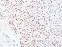
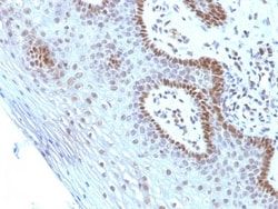
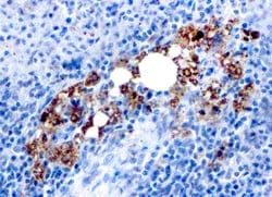
MITF (microphthalmia transcription factor) is a basic helix-loop-helix-leucine-zipper (bHLH-Zip) transcription factor that regulates the development and survival of melanocytes and retinal pigment epithelium, and also is involved in transcription of pigmentation enzyme genes such as tyrosinase TRP1 and TRP2. MITF has been shown to be phosphorylated by MAP kinase in response to c-kit activation, resulting in upregulation of MITF transcriptional activity. Mutations of the MITF gene are associated with the autosomal dominant hereditary deafness and pigmentation condition, Waardenburg Syndrome type 2A. Multiple isoforms of MITF exist, including MITF-A, MITF-B, MITF-C, MITF-H, and MITF-M, which differ in the amino-terminal domain and in their expression patterns. The MITF-M isoform is restricted to the melanocyte cell lineage. Anti-MITF, D5, recognizes a nuclear protein, which is expressed in the majority of primary and metastatic epithelioid malignant melanomas as well as in normal melanoc
Content And Storage
Store at 4C.
Isotype
IgG1 κ
Related Products

Description
- Ensure accurate, reproducible results in Flow Cytometry, Immunohistochemistry (Paraffin), Immunofluorescence MITF Monoclonal specifically detects MITF in Human, Mouse (Negative), Rat (Negative) samples
- It is validated for Immunohistochemistry, Immunohistochemistry-Paraffin.
Compare Similar Items
Show Difference
Antigen: MITF
Classification: Monoclonal
Concentration: 0.2mg/mL
Dilution: Flow Cytometry 0.5 - 1 ug/million cells in 0.1 ml, Immunohistochemistry-Paraffin 0.5 - 1.0 ug/ml, SDS-Page, Immunofluorescence 0.5 - 1.0 ug/ml
Gene Alias: BHLHE32, bHLHe32MI, Class E basic helix-loop-helix protein 32, microphthalmia-associated transcription factor, Waardenburg syndrome, type 2A, WS2A
Host Species: Mouse
Purification Method: Protein A or G purified
Regulatory Status: RUO
Gene ID (Entrez): 4286
Target Species: Human, Mouse (Negative), Rat (Negative)
Form: Purified
Applications: Flow Cytometry, Immunohistochemistry (Paraffin), SDS-Page, Immunofluorescence
Clone: D5
Conjugate: Unconjugated
Formulation: 10mM PBS and 0.05% BSA with 0.05% Sodium Azide
Gene Symbols: MITF
Immunogen: NH2 terminus fragment of human Mi protein
Quantity: 0.02 mg
Primary or Secondary: Primary
Test Specificity: MITF (microphthalmia transcription factor) is a basic helix-loop-helix-leucine-zipper (bHLH-Zip) transcription factor that regulates the development and survival of melanocytes and retinal pigment epithelium, and also is involved in transcription of pigmentation enzyme genes such as tyrosinase TRP1 and TRP2. MITF has been shown to be phosphorylated by MAP kinase in response to c-kit activation, resulting in upregulation of MITF transcriptional activity. Mutations of the MITF gene are associated with the autosomal dominant hereditary deafness and pigmentation condition, Waardenburg Syndrome type 2A. Multiple isoforms of MITF exist, including MITF-A, MITF-B, MITF-C, MITF-H, and MITF-M, which differ in the amino-terminal domain and in their expression patterns. The MITF-M isoform is restricted to the melanocyte cell lineage. Anti-MITF, D5, recognizes a nuclear protein, which is expressed in the majority of primary and metastatic epithelioid malignant melanomas as well as in normal melanoc
Content And Storage: Store at 4C.
Isotype: IgG1 κ
Antigen: MITF
Classification: Monoclonal
Concentration: 0.2mg/mL
Dilution: Flow Cytometry 0.5 - 1 ug/million cells in 0.1 ml, Immunohistochemistry-Paraffin 0.5 - 1.0 ug/ml, SDS-Page, Immunofluorescence 0.5 - 1.0 ug/ml
Gene Alias: BHLHE32, bHLHe32MI, Class E basic helix-loop-helix protein 32, microphthalmia-associated transcription factor, Waardenburg syndrome, type 2A, WS2A
Host Species: Mouse
Purification Method: Protein A or G purified
Regulatory Status: RUO
Gene ID (Entrez): 4286
Target Species: Human, Mouse (Negative), Rat (Negative)
Form: Purified
Applications: Flow Cytometry, Immunohistochemistry (Paraffin), SDS-Page, Immunofluorescence
Clone: D5
Conjugate: Unconjugated
Formulation: 10mM PBS and 0.05% BSA with 0.05% Sodium Azide
Gene Symbols: MITF
Immunogen: NH2 terminus fragment of human Mi protein
Quantity: 0.1 mg
Primary or Secondary: Primary
Test Specificity: MITF (microphthalmia transcription factor) is a basic helix-loop-helix-leucine-zipper (bHLH-Zip) transcription factor that regulates the development and survival of melanocytes and retinal pigment epithelium, and also is involved in transcription of pigmentation enzyme genes such as tyrosinase TRP1 and TRP2. MITF has been shown to be phosphorylated by MAP kinase in response to c-kit activation, resulting in upregulation of MITF transcriptional activity. Mutations of the MITF gene are associated with the autosomal dominant hereditary deafness and pigmentation condition, Waardenburg Syndrome type 2A. Multiple isoforms of MITF exist, including MITF-A, MITF-B, MITF-C, MITF-H, and MITF-M, which differ in the amino-terminal domain and in their expression patterns. The MITF-M isoform is restricted to the melanocyte cell lineage. Anti-MITF, D5, recognizes a nuclear protein, which is expressed in the majority of primary and metastatic epithelioid malignant melanomas as well as in normal melanoc
Content And Storage: Store at 4C.
Isotype: IgG1 κ
Antigen: MITF
Classification: Monoclonal
Concentration: 0.2mg/mL
Dilution: Flow Cytometry 0.5 - 1 ug/million cells in 0.1 ml, Immunohistochemistry-Paraffin 0.5 - 1.0 ug/ml, SDS-Page, Immunofluorescence 0.5 - 1.0 ug/ml
Gene Alias: BHLHE32, bHLHe32MI, Class E basic helix-loop-helix protein 32, microphthalmia-associated transcription factor, Waardenburg syndrome, type 2A, WS2A
Host Species: Mouse
Purification Method: Protein A or G purified
Regulatory Status: RUO
Gene ID (Entrez): 4286
Target Species: Human, Mouse (Negative), Rat (Negative)
Form: Purified
Applications: Flow Cytometry, Immunohistochemistry (Paraffin), SDS-Page, Immunofluorescence
Clone: D5
Conjugate: Unconjugated
Formulation: 10mM PBS and 0.05% BSA with 0.05% Sodium Azide
Gene Symbols: MITF
Immunogen: NH2 terminus fragment of human Mi protein
Quantity: 0.2 mg
Primary or Secondary: Primary
Test Specificity: MITF (microphthalmia transcription factor) is a basic helix-loop-helix-leucine-zipper (bHLH-Zip) transcription factor that regulates the development and survival of melanocytes and retinal pigment epithelium, and also is involved in transcription of pigmentation enzyme genes such as tyrosinase TRP1 and TRP2. MITF has been shown to be phosphorylated by MAP kinase in response to c-kit activation, resulting in upregulation of MITF transcriptional activity. Mutations of the MITF gene are associated with the autosomal dominant hereditary deafness and pigmentation condition, Waardenburg Syndrome type 2A. Multiple isoforms of MITF exist, including MITF-A, MITF-B, MITF-C, MITF-H, and MITF-M, which differ in the amino-terminal domain and in their expression patterns. The MITF-M isoform is restricted to the melanocyte cell lineage. Anti-MITF, D5, recognizes a nuclear protein, which is expressed in the majority of primary and metastatic epithelioid malignant melanomas as well as in normal melanoc
Content And Storage: Store at 4C.
Isotype: IgG1 κ