NBP24522700

NuMA Antibody (SPM300), Novus Biologicals™

Manufacturer: Fischer Scientific

Select a Size

Pack Size SKU Availability Price
Each of 1 NBP24522700-Each-of-1 In Stock ₹ 22,673.40

NBP24522700 - Each of 1

₹ 22,673.40

In Stock

Quantity

1

Base Price: ₹ 22,673.40

GST (18%): ₹ 4,081.212

Total Price: ₹ 26,754.612

Antigen

NuMA

Classification

Monoclonal

Concentration

0.2 mg/mL

Dilution

Flow Cytometry 0.5 - 1 ug/million cells in 0.1 ml, Immunohistochemistry-Paraffin 0.5 - 1.0 ug/ml, Immunofluorescence 0.5 - 1.0 ug/ml

Gene Alias

centrophilin stabilizes mitotic spindle in mitotic cells, nuclear mitotic apparatus protein 1, NUMA, NuMA protein, SP-H antigen, structural nuclear protein

Host Species

Mouse

Molecular Weight of Antigen

228 kDa

Quantity

0.02 mg

Research Discipline

Breast Cancer, Cell Biology, Cell Cycle and Replication, Cellular Markers, Mitotic Regulators

Gene ID (Entrez)

4926

Target Species

Human

Form

Purified

Applications

Flow Cytometry, Immunohistochemistry (Paraffin), Immunofluorescence

Clone

SPM300

Conjugate

Unconjugated

Formulation

10mM PBS and 0.05% BSA with 0.05% Sodium Azide

Gene Symbols

NUMA1

Immunogen

Colon carcinoma 174T cells

Purification Method

Protein A or G purified

Regulatory Status

RUO

Primary or Secondary

Primary

Test Specificity

Recognizes a phosphorylated protein of 228kDa, identified as nuclear mitotic apparatus protein (NuMA). Its epitope is resistant to phosphatases. NuMA is intra-nuclear protein and present in nucleus during interphase. At the onset of mitosis, it redistributes from the nucleus to two centrosomal structures that later will become part of the mitotic spindle pole. After anaphase, the protein redistributes from the spindle polar region into reforming nucleus. NuMA is an essential protein during mitosis for the terminal phases of chromosome separation and/or nuclear reassembly. Recently a study shows that NuMA is cleaved to a 180 to 200kDa during apoptosis. Chromosomal translocation of this gene with the RARA (retinoic acid receptor, alpha) gene on chromosome 17 has been detected in patients with acute promyelocytic leukemia.

Content And Storage

Store at 4C.

Isotype

IgM κ

Related Products

Img

Fischer Scientific

NBP24522702

--

Img

Fischer Scientific

NBP24522701

--

Img

Fischer Scientific

NBP24529400

--

Img

Fischer Scientific

NBP24529402

--

Img

Fischer Scientific

NBP24529401

--

Img

Fischer Scientific

NBP24529300

--

Img

Fischer Scientific

NBP24529302

--

Img

Fischer Scientific

NBP24529301

--

Description

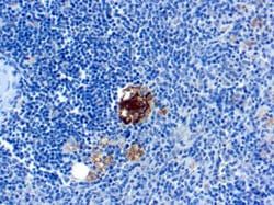
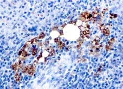
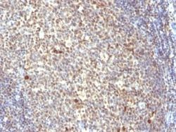
  • Ensure accurate, reproducible results in Flow Cytometry, Immunohistochemistry (Paraffin), Immunofluorescence NuMA Monoclonal specifically detects NuMA in Human samples
  • It is validated for Flow Cytometry, Immunohistochemistry, Immunocytochemistry/Immunofluorescence, Immunohistochemistry-Paraffin, Immunofluorescence.
Compare Similar Items

Show Difference

Img

Fischer Scientific

NBP24522700

--


Antigen:
NuMA

Classification:
Monoclonal

Concentration:
0.2 mg/mL

Dilution:
Flow Cytometry 0.5 - 1 ug/million cells in 0.1 ml, Immunohistochemistry-Paraffin 0.5 - 1.0 ug/ml, Immunofluorescence 0.5 - 1.0 ug/ml

Gene Alias:
centrophilin stabilizes mitotic spindle in mitotic cells, nuclear mitotic apparatus protein 1, NUMA, NuMA protein, SP-H antigen, structural nuclear protein

Host Species:
Mouse

Molecular Weight of Antigen:
228 kDa

Quantity:
0.02 mg

Research Discipline:
Breast Cancer, Cell Biology, Cell Cycle and Replication, Cellular Markers, Mitotic Regulators

Gene ID (Entrez):
4926

Target Species:
Human

Form:
Purified

Applications:
Flow Cytometry, Immunohistochemistry (Paraffin), Immunofluorescence

Clone:
SPM300

Conjugate:
Unconjugated

Formulation:
10mM PBS and 0.05% BSA with 0.05% Sodium Azide

Gene Symbols:
NUMA1

Immunogen:
Colon carcinoma 174T cells

Purification Method:
Protein A or G purified

Regulatory Status:
RUO

Primary or Secondary:
Primary

Test Specificity:
Recognizes a phosphorylated protein of 228kDa, identified as nuclear mitotic apparatus protein (NuMA). Its epitope is resistant to phosphatases. NuMA is intra-nuclear protein and present in nucleus during interphase. At the onset of mitosis, it redistributes from the nucleus to two centrosomal structures that later will become part of the mitotic spindle pole. After anaphase, the protein redistributes from the spindle polar region into reforming nucleus. NuMA is an essential protein during mitosis for the terminal phases of chromosome separation and/or nuclear reassembly. Recently a study shows that NuMA is cleaved to a 180 to 200kDa during apoptosis. Chromosomal translocation of this gene with the RARA (retinoic acid receptor, alpha) gene on chromosome 17 has been detected in patients with acute promyelocytic leukemia.

Content And Storage:
Store at 4C.

Isotype:
IgM κ

Img

Fischer Scientific

NBP24522701

--


Antigen:
NuMA

Classification:
Monoclonal

Concentration:
0.2 mg/mL

Dilution:
Flow Cytometry 0.5 - 1 ug/million cells in 0.1 ml, Immunohistochemistry-Paraffin 0.5 - 1.0 ug/ml, Immunofluorescence 0.5 - 1.0 ug/ml

Gene Alias:
centrophilin stabilizes mitotic spindle in mitotic cells, nuclear mitotic apparatus protein 1, NUMA, NuMA protein, SP-H antigen, structural nuclear protein

Host Species:
Mouse

Molecular Weight of Antigen:
228 kDa

Quantity:
0.1 mg

Research Discipline:
Breast Cancer, Cell Biology, Cell Cycle and Replication, Cellular Markers, Mitotic Regulators

Gene ID (Entrez):
4926

Target Species:
Human

Form:
Purified

Applications:
Flow Cytometry, Immunohistochemistry (Paraffin), Immunofluorescence

Clone:
SPM300

Conjugate:
Unconjugated

Formulation:
10mM PBS and 0.05% BSA with 0.05% Sodium Azide

Gene Symbols:
NUMA1

Immunogen:
Colon carcinoma 174T cells

Purification Method:
Protein A or G purified

Regulatory Status:
RUO

Primary or Secondary:
Primary

Test Specificity:
Recognizes a phosphorylated protein of 228kDa, identified as nuclear mitotic apparatus protein (NuMA). Its epitope is resistant to phosphatases. NuMA is intra-nuclear protein and present in nucleus during interphase. At the onset of mitosis, it redistributes from the nucleus to two centrosomal structures that later will become part of the mitotic spindle pole. After anaphase, the protein redistributes from the spindle polar region into reforming nucleus. NuMA is an essential protein during mitosis for the terminal phases of chromosome separation and/or nuclear reassembly. Recently a study shows that NuMA is cleaved to a 180 to 200kDa during apoptosis. Chromosomal translocation of this gene with the RARA (retinoic acid receptor, alpha) gene on chromosome 17 has been detected in patients with acute promyelocytic leukemia.

Content And Storage:
Store at 4C.

Isotype:
IgM κ

Img

Fischer Scientific

NBP24522702

--


Antigen:
NuMA

Classification:
Monoclonal

Concentration:
0.2 mg/mL

Dilution:
Flow Cytometry 0.5 - 1 ug/million cells in 0.1 ml, Immunohistochemistry-Paraffin 0.5 - 1.0 ug/ml, Immunofluorescence 0.5 - 1.0 ug/ml

Gene Alias:
centrophilin stabilizes mitotic spindle in mitotic cells, nuclear mitotic apparatus protein 1, NUMA, NuMA protein, SP-H antigen, structural nuclear protein

Host Species:
Mouse

Molecular Weight of Antigen:
228 kDa

Quantity:
0.2 mg

Research Discipline:
Breast Cancer, Cell Biology, Cell Cycle and Replication, Cellular Markers, Mitotic Regulators

Gene ID (Entrez):
4926

Target Species:
Human

Form:
Purified

Applications:
Flow Cytometry, Immunohistochemistry (Paraffin), Immunofluorescence

Clone:
SPM300

Conjugate:
Unconjugated

Formulation:
10mM PBS and 0.05% BSA with 0.05% Sodium Azide

Gene Symbols:
NUMA1

Immunogen:
Colon carcinoma 174T cells

Purification Method:
Protein A or G purified

Regulatory Status:
RUO

Primary or Secondary:
Primary

Test Specificity:
Recognizes a phosphorylated protein of 228kDa, identified as nuclear mitotic apparatus protein (NuMA). Its epitope is resistant to phosphatases. NuMA is intra-nuclear protein and present in nucleus during interphase. At the onset of mitosis, it redistributes from the nucleus to two centrosomal structures that later will become part of the mitotic spindle pole. After anaphase, the protein redistributes from the spindle polar region into reforming nucleus. NuMA is an essential protein during mitosis for the terminal phases of chromosome separation and/or nuclear reassembly. Recently a study shows that NuMA is cleaved to a 180 to 200kDa during apoptosis. Chromosomal translocation of this gene with the RARA (retinoic acid receptor, alpha) gene on chromosome 17 has been detected in patients with acute promyelocytic leukemia.

Content And Storage:
Store at 4C.

Isotype:
IgM κ

Img

Fischer Scientific

NBP24523000

--


Antigen:
CD90/Thy1

Classification:
Monoclonal

Concentration:
0.2 mg/mL

Dilution:
Flow Cytometry 0.5 - 1 ug/million cells in 0.1 ml, SDS-Page, Immunofluorescence 0.5 - 1.0 ug/ml

Gene Alias:
CD90, CD90 antigen, CDw90, FLJ33325, Thy-1 antigen, Thy-1 cell surface antigen, thy-1 membrane glycoprotein, Thy-1 T-cell antigen

Host Species:
Mouse

Molecular Weight of Antigen:
__

Quantity:
0.02 mg

Research Discipline:
Hematopoietic Stem Cell Markers, Hepatic Stem Cell Markers, Immunology, Innate Immunity, Mesenchymal Stem Cell Markers, Signal Transduction, Stem Cell Markers

Gene ID (Entrez):
7070

Target Species:
Human, Bovine

Form:
Purified

Applications:
Flow Cytometry, SDS-Page, Immunofluorescence

Clone:
AF-9

Conjugate:
Unconjugated

Formulation:
10mM PBS and 0.05% BSA with 0.05% Sodium Azide

Gene Symbols:
THY1

Immunogen:
Human T-lymphoma cells

Purification Method:
Protein A or G purified

Regulatory Status:
RUO

Primary or Secondary:
Primary

Test Specificity:
Recognizes a protein of 18-35kDa, identified as CD90 (also known as Thy1). CD90 is a member of the immunoglobulin superfamily. It may contribute to inhibition of proliferation/differentiation of hematopoietic stem cells and neuron memory formation in the CNS. It consists of a single Ig domain (112 amino acids; 25-35 kDa) inserted into the cell membrane via a GPI anchor. Expressed by hematopoietic stem cells and neurons in all species studied. Its highly expressed in connective tissue and various fibroblast and stromal cell lines, expressed on all thymocytes and peripheral T cells in mice, but in humans expressed only on small % fetal thymocytes, 10-40% of CD34+ cells in bone marrow, and <1% of CD3+CD4+ lymphocytes in peripheral circulation. It is also expressed by human lymph node HEV endothelium but not other endothelia. Lastly, it is expressed by a limited number of lymphoblastoid and leukemic cell lines.

Content And Storage:
Store at 4C.

Isotype:
IgG1 κ