Lewis A Blood Group Antigen Antibody (SPM279), Novus Biologicals™
Manufacturer: Fischer Scientific
Select a Size
| Pack Size | SKU | Availability | Price |
|---|---|---|---|
| Each of 1 | NBP24528300-Each-of-1 | In Stock | ₹ 23,852.00 |
NBP24528300 - Each of 1
In Stock
Quantity
1
Base Price: ₹ 23,852.00
GST (18%): ₹ 4,293.36
Total Price: ₹ 28,145.36
Antigen
Lewis A Blood Group Antigen
Classification
Monoclonal
Concentration
0.2mg/mL
Dilution
Flow Cytometry 0.5 - 1 ug/million cells in 0.1 ml, Immunohistochemistry-Paraffin 0.5 - 1.0 ug/ml, Immunofluorescence 0.5 - 1.0 ug/ml
Gene Symbols
ABO
Immunogen
Mucins isolated from ovarian cyst fluid
Quantity
0.02 mg
Primary or Secondary
Primary
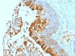
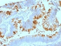
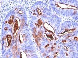
Test Specificity
Recognizes a carbohydrate determinant of Gal 1-3(Fuc 1-4) GlcNAc which is blood group antigen Lewis A. It is present primarily on epithelial cells such as colon and kidneys. In the tumors and dedifferentiated tissues, decrease of Lewis A antigen was observed. Lewis A (type 1 chain) is expressed in colonic epithelial cells and may be useful for detection of gastrointestinal tumors, pancreatic cancer, and colorectal tumors. Blood group related antigens represent a group of carbohydrate determinants carried on both glycolipids and glycoproteins. They are usually mucin-type, and are detected on erythrocytes, certain epithelial cells, and in secretions of certain individuals. Sixteen genetically and biosynthetically distinct but inter-related specificities belong to this group of antigens, including A, B, H, Lewis A, Lewis B, Lewis X, Lewis Y, and precursor type 1 chain antigens.
Content And Storage
Store at 4C.
Isotype
IgG1 κ
Applications
Flow Cytometry, Immunohistochemistry (Paraffin), Immunofluorescence
Clone
SPM279
Conjugate
Unconjugated
Formulation
10mM PBS and 0.05% BSA with 0.05% Sodium Azide
Host Species
Mouse
Purification Method
Protein A or G purified
Regulatory Status
RUO
Gene ID (Entrez)
28
Target Species
Human, Mouse
Form
Purified
Related Products

Description
- Ensure accurate, reproducible results in Flow Cytometry, Immunohistochemistry (Paraffin), Immunofluorescence Lewis A Blood Group Antigen Monoclonal specifically detects Lewis A Blood Group Antigen in Human, Mouse samples
- It is validated for Flow Cytometry, Immunohistochemistry, Immunocytochemistry/Immunofluorescence, Immunohistochemistry-Paraffin, Immunofluorescence.
Compare Similar Items
Show Difference
Antigen: Lewis A Blood Group Antigen
Classification: Monoclonal
Concentration: 0.2mg/mL
Dilution: Flow Cytometry 0.5 - 1 ug/million cells in 0.1 ml, Immunohistochemistry-Paraffin 0.5 - 1.0 ug/ml, Immunofluorescence 0.5 - 1.0 ug/ml
Gene Symbols: ABO
Immunogen: Mucins isolated from ovarian cyst fluid
Quantity: 0.02 mg
Primary or Secondary: Primary
Test Specificity: Recognizes a carbohydrate determinant of Gal 1-3(Fuc 1-4) GlcNAc which is blood group antigen Lewis A. It is present primarily on epithelial cells such as colon and kidneys. In the tumors and dedifferentiated tissues, decrease of Lewis A antigen was observed. Lewis A (type 1 chain) is expressed in colonic epithelial cells and may be useful for detection of gastrointestinal tumors, pancreatic cancer, and colorectal tumors. Blood group related antigens represent a group of carbohydrate determinants carried on both glycolipids and glycoproteins. They are usually mucin-type, and are detected on erythrocytes, certain epithelial cells, and in secretions of certain individuals. Sixteen genetically and biosynthetically distinct but inter-related specificities belong to this group of antigens, including A, B, H, Lewis A, Lewis B, Lewis X, Lewis Y, and precursor type 1 chain antigens.
Content And Storage: Store at 4C.
Isotype: IgG1 κ
Applications: Flow Cytometry, Immunohistochemistry (Paraffin), Immunofluorescence
Clone: SPM279
Conjugate: Unconjugated
Formulation: 10mM PBS and 0.05% BSA with 0.05% Sodium Azide
Host Species: Mouse
Purification Method: Protein A or G purified
Regulatory Status: RUO
Gene ID (Entrez): 28
Target Species: Human, Mouse
Form: Purified
Antigen: Lewis A Blood Group Antigen
Classification: Monoclonal
Concentration: 0.2mg/mL
Dilution: Flow Cytometry 0.5 - 1 ug/million cells in 0.1 ml, Immunohistochemistry-Paraffin 0.5 - 1.0 ug/ml, Immunofluorescence 0.5 - 1.0 ug/ml
Gene Symbols: ABO
Immunogen: Mucins isolated from ovarian cyst fluid
Quantity: 0.1 mg
Primary or Secondary: Primary
Test Specificity: Recognizes a carbohydrate determinant of Gal 1-3(Fuc 1-4) GlcNAc which is blood group antigen Lewis A. It is present primarily on epithelial cells such as colon and kidneys. In the tumors and dedifferentiated tissues, decrease of Lewis A antigen was observed. Lewis A (type 1 chain) is expressed in colonic epithelial cells and may be useful for detection of gastrointestinal tumors, pancreatic cancer, and colorectal tumors. Blood group related antigens represent a group of carbohydrate determinants carried on both glycolipids and glycoproteins. They are usually mucin-type, and are detected on erythrocytes, certain epithelial cells, and in secretions of certain individuals. Sixteen genetically and biosynthetically distinct but inter-related specificities belong to this group of antigens, including A, B, H, Lewis A, Lewis B, Lewis X, Lewis Y, and precursor type 1 chain antigens.
Content And Storage: Store at 4C.
Isotype: IgG1 κ
Applications: Flow Cytometry, Immunohistochemistry (Paraffin), Immunofluorescence
Clone: SPM279
Conjugate: Unconjugated
Formulation: 10mM PBS and 0.05% BSA with 0.05% Sodium Azide
Host Species: Mouse
Purification Method: Protein A or G purified
Regulatory Status: RUO
Gene ID (Entrez): 28
Target Species: Human, Mouse
Form: Purified
Antigen: Lewis A Blood Group Antigen
Classification: Monoclonal
Concentration: 0.2mg/mL
Dilution: Flow Cytometry 0.5 - 1 ug/million cells in 0.1 ml, Immunohistochemistry-Paraffin 0.5 - 1.0 ug/ml, Immunofluorescence 0.5 - 1.0 ug/ml
Gene Symbols: ABO
Immunogen: Mucins isolated from ovarian cyst fluid
Quantity: 0.2 mg
Primary or Secondary: Primary
Test Specificity: Recognizes a carbohydrate determinant of Gal 1-3(Fuc 1-4) GlcNAc which is blood group antigen Lewis A. It is present primarily on epithelial cells such as colon and kidneys. In the tumors and dedifferentiated tissues, decrease of Lewis A antigen was observed. Lewis A (type 1 chain) is expressed in colonic epithelial cells and may be useful for detection of gastrointestinal tumors, pancreatic cancer, and colorectal tumors. Blood group related antigens represent a group of carbohydrate determinants carried on both glycolipids and glycoproteins. They are usually mucin-type, and are detected on erythrocytes, certain epithelial cells, and in secretions of certain individuals. Sixteen genetically and biosynthetically distinct but inter-related specificities belong to this group of antigens, including A, B, H, Lewis A, Lewis B, Lewis X, Lewis Y, and precursor type 1 chain antigens.
Content And Storage: Store at 4C.
Isotype: IgG1 κ
Applications: Flow Cytometry, Immunohistochemistry (Paraffin), Immunofluorescence
Clone: SPM279
Conjugate: Unconjugated
Formulation: 10mM PBS and 0.05% BSA with 0.05% Sodium Azide
Host Species: Mouse
Purification Method: Protein A or G purified
Regulatory Status: RUO
Gene ID (Entrez): 28
Target Species: Human, Mouse
Form: Purified