Melanoma Associated Antigen (PNL2) Antibody (PNL2), Novus Biologicals™
Manufacturer: Fischer Scientific
Select a Size
| Pack Size | SKU | Availability | Price |
|---|---|---|---|
| Each of 1 | NBP24528401-Each-of-1 | In Stock | ₹ 46,636.00 |
NBP24528401 - Each of 1
In Stock
Quantity
1
Base Price: ₹ 46,636.00
GST (18%): ₹ 8,394.48
Total Price: ₹ 55,030.48
Antigen
Melanoma Associated Antigen (PNL2)
Classification
Monoclonal
Concentration
0.2mg/mL
Dilution
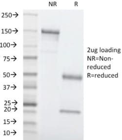
Immunohistochemistry-Paraffin 0.5 - 1.0 ug/ml, SDS-Page, Immunofluorescence 0.5 - 1.0 ug/ml
Host Species
Mouse
Purification Method
Protein A or G purified
Regulatory Status
RUO
Test Specificity
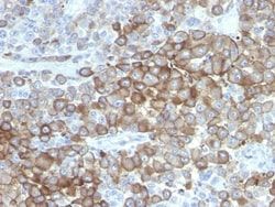
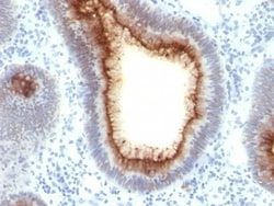
Anti-PNL2 is a novel monoclonal antibody, which has recently been introduced as an immunohistochemical reagent to stain melanocytes and tumors derived therefrom. The antigen recognized by PNL2 is different from Melan A and gp100. Its epitope is not destroyed by digestion with neuraminidase i.e. its epitope id not glycosylated. Anti-PNL2 may be most useful because of its high sensitivity for metastatic melanoma (87%), as opposed to 76% for anti-HMB45 and 82% for anti-MART-1. Anti-PNL2 labels intra-epidermal nevi while the dermal component of compound nevi are largely non-reactive with anti-PNL2. Antibodies against PNL2, MART-1 (Melan A) and HMB45 stain most clear cell sarcoma cells and a few cells in angio-myolipomas and lymphangioleiomyomatosis. Anti-PNL2 is a useful antibody for the identification of melanomas and clear cell sarcomas. Differential diagnosis is aided by the results from a panel of antibodies, including antibodies against HMB45, MART-1, tyrosinase, and MiTF.
Content And Storage
Store at 4C.
Isotype
IgG1
Applications
Immunohistochemistry (Paraffin), SDS-Page, Immunofluorescence
Clone
PNL2
Conjugate
Unconjugated
Formulation
10mM PBS and 0.05% BSA with 0.05% Sodium Azide
Immunogen
Melanocyte antigen
Quantity
0.1 mg
Primary or Secondary
Primary
Target Species
Human, Mouse, Canine
Form
Purified
Related Products

Description
- Ensure accurate, reproducible results in Immunohistochemistry (Paraffin), Immunofluorescence Melanoma Associated Antigen (PNL2) Monoclonal specifically detects Melanoma Associated Antigen (PNL2) in Human, Canine samples
- It is validated for Immunohistochemistry, Immunocytochemistry/Immunofluorescence, Immunohistochemistry-Paraffin, Immunofluorescence.
Compare Similar Items
Show Difference
Antigen: Melanoma Associated Antigen (PNL2)
Classification: Monoclonal
Concentration: 0.2mg/mL
Dilution: Immunohistochemistry-Paraffin 0.5 - 1.0 ug/ml, SDS-Page, Immunofluorescence 0.5 - 1.0 ug/ml
Host Species: Mouse
Purification Method: Protein A or G purified
Regulatory Status: RUO
Test Specificity: Anti-PNL2 is a novel monoclonal antibody, which has recently been introduced as an immunohistochemical reagent to stain melanocytes and tumors derived therefrom. The antigen recognized by PNL2 is different from Melan A and gp100. Its epitope is not destroyed by digestion with neuraminidase i.e. its epitope id not glycosylated. Anti-PNL2 may be most useful because of its high sensitivity for metastatic melanoma (87%), as opposed to 76% for anti-HMB45 and 82% for anti-MART-1. Anti-PNL2 labels intra-epidermal nevi while the dermal component of compound nevi are largely non-reactive with anti-PNL2. Antibodies against PNL2, MART-1 (Melan A) and HMB45 stain most clear cell sarcoma cells and a few cells in angio-myolipomas and lymphangioleiomyomatosis. Anti-PNL2 is a useful antibody for the identification of melanomas and clear cell sarcomas. Differential diagnosis is aided by the results from a panel of antibodies, including antibodies against HMB45, MART-1, tyrosinase, and MiTF.
Content And Storage: Store at 4C.
Isotype: IgG1
Applications: Immunohistochemistry (Paraffin), SDS-Page, Immunofluorescence
Clone: PNL2
Conjugate: Unconjugated
Formulation: 10mM PBS and 0.05% BSA with 0.05% Sodium Azide
Immunogen: Melanocyte antigen
Quantity: 0.1 mg
Primary or Secondary: Primary
Target Species: Human, Mouse, Canine
Form: Purified
Antigen: Melanoma Associated Antigen (PNL2)
Classification: Monoclonal
Concentration: 0.2mg/mL
Dilution: Immunohistochemistry-Paraffin 0.5 - 1.0 ug/ml, SDS-Page, Immunofluorescence 0.5 - 1.0 ug/ml
Host Species: Mouse
Purification Method: Protein A or G purified
Regulatory Status: RUO
Test Specificity: Anti-PNL2 is a novel monoclonal antibody, which has recently been introduced as an immunohistochemical reagent to stain melanocytes and tumors derived therefrom. The antigen recognized by PNL2 is different from Melan A and gp100. Its epitope is not destroyed by digestion with neuraminidase i.e. its epitope id not glycosylated. Anti-PNL2 may be most useful because of its high sensitivity for metastatic melanoma (87%), as opposed to 76% for anti-HMB45 and 82% for anti-MART-1. Anti-PNL2 labels intra-epidermal nevi while the dermal component of compound nevi are largely non-reactive with anti-PNL2. Antibodies against PNL2, MART-1 (Melan A) and HMB45 stain most clear cell sarcoma cells and a few cells in angio-myolipomas and lymphangioleiomyomatosis. Anti-PNL2 is a useful antibody for the identification of melanomas and clear cell sarcomas. Differential diagnosis is aided by the results from a panel of antibodies, including antibodies against HMB45, MART-1, tyrosinase, and MiTF.
Content And Storage: Store at 4C.
Isotype: IgG1
Applications: Immunohistochemistry (Paraffin), SDS-Page, Immunofluorescence
Clone: PNL2
Conjugate: Unconjugated
Formulation: 10mM PBS and 0.05% BSA with 0.05% Sodium Azide
Immunogen: Melanocyte antigen
Quantity: 0.2 mg
Primary or Secondary: Primary
Target Species: Human, Mouse, Canine
Form: Purified
Antigen: S100A9
Classification: Monoclonal
Concentration: 0.2 mg/mL
Dilution: Flow Cytometry 0.5 - 1 ug/million cells in 0.1 ml, Immunohistochemistry-Paraffin 0.5 - 1 ug/ml, Immunohistochemistry-Frozen (Negative), Immunofluorescence 0.5 - 1.0 ug/ml
Host Species: Mouse
Purification Method: Protein A or G purified
Regulatory Status: RUO
Test Specificity: Recognizes a protein of14kDa, identified as MRP-14 (also known as Calgranulin B or S100AA9). It comprises 60% of the cytoplasmic protein fraction of circulating polymorphonuclear granulocytes and is also found in monocytes, macrophages and ileal tissue eosinophils. Peripheral blood monocytes carry the antigen extra- and intracellularly, neutrophils only intracellularly. It is a potent chemotactic factor for neutrophils. Plasma concentrations are elevated in diseases associated with increased neutrophil activity, like inflammatory bowel disease. Granulocytes terminate their existence after transmigration through the intestinal wall. Therefore, it is also detectable in feces. Elevated levels have been observed in body fluids such as plasma, saliva, gingival crevicular fluid, stools, and synovial fluid during infection and inflammatory conditions. This MAb reacts with neutrophils, monocytes, and macrophages, and has been shown as an important marker for identifying macrophages in tissue s
Content And Storage: Store at 4C.
Isotype: IgM κ
Applications: Flow Cytometry, Immunohistochemistry (Paraffin), Immunohistochemistry (Frozen), Immunofluorescence
Clone: MRP14/840
Conjugate: Unconjugated
Formulation: 1.0mM PBS and 0.05% BSA with 0.05% Sodium Azide
Immunogen: Recombinant human MRP14 protein
Quantity: 0.02 mg
Primary or Secondary: Primary
Target Species: Human
Form: Purified