MLH1 Mouse anti-Human, Clone: MLH1/1324, Novus Biologicals™
Manufacturer: Fischer Scientific
Select a Size
| Pack Size | SKU | Availability | Price |
|---|---|---|---|
| Each of 1 | NBP247656A-Each-of-1 | In Stock | ₹ 43,432.00 |
NBP247656A - Each of 1
In Stock
Quantity
1
Base Price: ₹ 43,432.00
GST (18%): ₹ 7,817.76
Total Price: ₹ 51,249.76
Antigen
MLH1
Classification
Monoclonal
Conjugate
Unconjugated
Formulation
10mM PBS and 0.05% BSA with 0.05% Sodium Azide
Gene Symbols
MLH1
Immunogen
Recombinant human MLH1 protein
Purification Method
Protein A or G purified
Regulatory Status
RUO
Primary or Secondary
Primary
Test Specificity
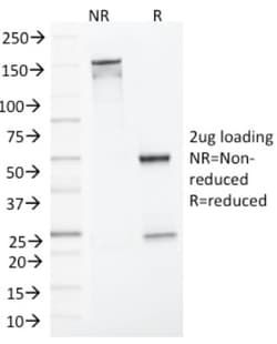
This MAb recognizes a protein of 83kDa, identified as MLH1. Its epitope maps in aa 380-410. Defects in MLH1 are the cause of hereditary non-polyposis colorectal cancer type 2 (HNPCC2). Heterodimerizes with PMS2 to form MutL alpha, a component of the post-replicative DNA mismatch repair system (MMR). DNA repair is initiated by MutS alpha (MSH2-MSH6) or MutS beta (MSH2-MSH6) binding to a dsDNA mismatch, then MutL alpha is recruited to the heteroduplex. Assembly of the MutL-MutS-heteroduplex ternary complex in presence of RFC and PCNA is sufficient to activate endonuclease activity of PMS2. It introduces single-strand breaks near the mismatch and thus generates new entry points for the exonuclease EXO1 to degrade the strand containing the mismatch. DNA methylation would prevent cleavage and therefore assure that only the newly mutated DNA strand is going to be corrected. MutL alpha (MLH1-PMS2) interacts physically with the clamp loader subunits of DNA polymerase III, suggesting that it ma
Content And Storage
Store at 4C.
Isotype
IgG2b
Applications
Western Blot, SDS-Page
Clone
MLH1/1324
Dilution
Western Blot 0.5-1.0ug/ml, SDS-Page
Gene Alias
COCA2FCC2, DNA mismatch repair protein Mlh1, hMLH1, HNPCC, HNPCC2MGC5172, mutL (E. coli) homolog 1 (colon cancer, nonpolyposis type 2), mutL homolog 1, colon cancer, nonpolyposis type 2 (E. coli), MutL protein homolog 1
Host Species
Mouse
Molecular Weight of Antigen
85 kDa
Quantity
0.1 mg
Research Discipline
Apoptosis, Breast Cancer, Cancer, Cell Cycle and Replication, Checkpoint signaling, DNA Double Strand Break Repair, DNA Repair, Mismatch Repair, Tumor Suppressors
Gene ID (Entrez)
4292
Target Species
Human
Form
Purified
Related Products

Description
- MLH1 Monoclonal specifically detects MLH1 in Human samples
- It is validated for ELISA.
Compare Similar Items
Show Difference
Antigen: MLH1
Classification: Monoclonal
Conjugate: Unconjugated
Formulation: 10mM PBS and 0.05% BSA with 0.05% Sodium Azide
Gene Symbols: MLH1
Immunogen: Recombinant human MLH1 protein
Purification Method: Protein A or G purified
Regulatory Status: RUO
Primary or Secondary: Primary
Test Specificity: This MAb recognizes a protein of 83kDa, identified as MLH1. Its epitope maps in aa 380-410. Defects in MLH1 are the cause of hereditary non-polyposis colorectal cancer type 2 (HNPCC2). Heterodimerizes with PMS2 to form MutL alpha, a component of the post-replicative DNA mismatch repair system (MMR). DNA repair is initiated by MutS alpha (MSH2-MSH6) or MutS beta (MSH2-MSH6) binding to a dsDNA mismatch, then MutL alpha is recruited to the heteroduplex. Assembly of the MutL-MutS-heteroduplex ternary complex in presence of RFC and PCNA is sufficient to activate endonuclease activity of PMS2. It introduces single-strand breaks near the mismatch and thus generates new entry points for the exonuclease EXO1 to degrade the strand containing the mismatch. DNA methylation would prevent cleavage and therefore assure that only the newly mutated DNA strand is going to be corrected. MutL alpha (MLH1-PMS2) interacts physically with the clamp loader subunits of DNA polymerase III, suggesting that it ma
Content And Storage: Store at 4C.
Isotype: IgG2b
Applications: Western Blot, SDS-Page
Clone: MLH1/1324
Dilution: Western Blot 0.5-1.0ug/ml, SDS-Page
Gene Alias: COCA2FCC2, DNA mismatch repair protein Mlh1, hMLH1, HNPCC, HNPCC2MGC5172, mutL (E. coli) homolog 1 (colon cancer, nonpolyposis type 2), mutL homolog 1, colon cancer, nonpolyposis type 2 (E. coli), MutL protein homolog 1
Host Species: Mouse
Molecular Weight of Antigen: 85 kDa
Quantity: 0.1 mg
Research Discipline: Apoptosis, Breast Cancer, Cancer, Cell Cycle and Replication, Checkpoint signaling, DNA Double Strand Break Repair, DNA Repair, Mismatch Repair, Tumor Suppressors
Gene ID (Entrez): 4292
Target Species: Human
Form: Purified
Antigen: MLH1
Classification: Monoclonal
Conjugate: Unconjugated
Formulation: 10mM PBS and 0.05% BSA with 0.05% Sodium Azide
Gene Symbols: MLH1
Immunogen: Recombinant human MLH1 protein
Purification Method: Protein A or G purified
Regulatory Status: RUO
Primary or Secondary: Primary
Test Specificity: This MAb recognizes a protein of 83kDa, identified as MLH1. Its epitope maps in aa 380-410. Defects in MLH1 are the cause of hereditary non-polyposis colorectal cancer type 2 (HNPCC2). Heterodimerizes with PMS2 to form MutL alpha, a component of the post-replicative DNA mismatch repair system (MMR). DNA repair is initiated by MutS alpha (MSH2-MSH6) or MutS beta (MSH2-MSH6) binding to a dsDNA mismatch, then MutL alpha is recruited to the heteroduplex. Assembly of the MutL-MutS-heteroduplex ternary complex in presence of RFC and PCNA is sufficient to activate endonuclease activity of PMS2. It introduces single-strand breaks near the mismatch and thus generates new entry points for the exonuclease EXO1 to degrade the strand containing the mismatch. DNA methylation would prevent cleavage and therefore assure that only the newly mutated DNA strand is going to be corrected. MutL alpha (MLH1-PMS2) interacts physically with the clamp loader subunits of DNA polymerase III, suggesting that it may play a role to recruit the DNA polymerase III to the site of the MMR. Also implicated in DNA damage signaling, a process, which induces cell cycle arrest and can lead to apoptosis in case of major DNA damages. Heterodimerizes with MLH3 to form MutL gamma, which plays a role in meiosis.
Content And Storage: Store at 4C.
Isotype: IgG2b
Applications: Western Blot, SDS-Page
Clone: MLH1/1324
Dilution: Western Blot 0.5-1.0ug/ml, SDS-Page
Gene Alias: COCA2FCC2, DNA mismatch repair protein Mlh1, hMLH1, HNPCC, HNPCC2MGC5172, mutL (E. coli) homolog 1 (colon cancer, nonpolyposis type 2), mutL homolog 1, colon cancer, nonpolyposis type 2 (E. coli), MutL protein homolog 1
Host Species: Mouse
Molecular Weight of Antigen: 85 kDa
Quantity: 0.2 mg
Research Discipline: Apoptosis, Breast Cancer, Cancer, Cell Cycle and Replication, Checkpoint signaling, DNA Double Strand Break Repair, DNA Repair, Mismatch Repair, Tumor Suppressors
Gene ID (Entrez): 4292
Target Species: Human
Form: Purified
Antigen: GPR119
Classification: Polyclonal
Conjugate: Unconjugated
Formulation: Tris 0,1M, glycine 0,1M, sucrose 2% with No Preservative
Gene Symbols: GPR119
Immunogen: A synthetic peptide derived from with the C-terminal domain of mouse Gpr119 protein.
Purification Method: Protein A purified
Regulatory Status: RUO
Primary or Secondary: Primary
Test Specificity: Reacts with mouse Gpr 119 protein. Cross reacts with rat protein due to sequence homology.
Content And Storage: Lyophilized antibody can be kept at 4C for up to 3 months and should be kept at -20C for long-term storage. To avoid freeze-thaw cycles, reconstituted antibody should be aliquoted before freezing for short-term storage (-20C) or for long-term storage (-80C). For maximum recovery of product, centrifuge the original vial prior to removing the cap. Further dilutions can be made in assay buffer.
Isotype: IgG
Applications: Western Blot, ELISA, Immunohistochemistry, Immunocytochemistry, Immunofluorescence
Clone: __
Dilution: Western Blot 1:1000 - 1:5000, ELISA 1:100 - 1:2000, Immunohistochemistry 1:100 - 1:000, Immunocytochemistry/Immunofluorescence 1:100-1:1000
Gene Alias: G protein-coupled receptor 119, glucose-dependent insulinotropic receptor, GPCR2, G-protein coupled receptor 119, G-protein coupled receptor 2, hGPCR2, MGC119957
Host Species: Rabbit
Molecular Weight of Antigen: __
Quantity: 0.1 mg
Research Discipline: GPCR
Gene ID (Entrez): 139760
Target Species: Mouse, Rat
Form: Purified
