TRACP/PAP/ACP5 Antibody (ACP5/1070) - Azide and BSA Free, Novus Biologicals™
Manufacturer: Fischer Scientific
The price for this product is unavailable. Please request a quote
Antigen
TRACP/PAP/ACP5
Classification
Monoclonal
Concentration
1.0 mg/mL
Dilution
Flow Cytometry : 0.5 - 1 ug/million cells in 0.1 ml, Immunohistochemistry-Paraffin : 0.5 - 1.0 ug/ml, Immunofluorescence : 0.5 - 1.0 ug/ml, CyTOF-ready
Gene Symbols
ACP5
Immunogen
Recombinant full-length human ACP5 protein
Purification Method
Protein A or G purified
Regulatory Status
RUO
Gene ID (Entrez)
54
Target Species
Human, Mouse, Rat
Form
Purified
Applications
Flow Cytometry, Immunohistochemistry (Paraffin), Immunofluorescence, CyTOF
Clone
ACP5/1070
Conjugate
Unconjugated
Formulation
PBS with No Preservative
Host Species
Mouse
Molecular Weight of Antigen
35 kDa
Quantity
0.2 mg
Primary or Secondary
Primary
Test Specificity
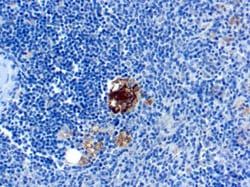
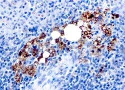
It recognizes a protein of 35kDa, which is identified as tartrate-resistant acid phosphatase (TRAcP). It exists as two isoforms (5a and 5b). This MAb reacts with both the isoforms. Serum TRAcP 5a is secreted by macrophages and dendritic cells and increased in many patients of rheumatoid arthritis.Serum TRAcP 5b is produced from osteoclasts and elevated during bone resorption. TRAcP is an iron containing glycoprotein, which catalyzes the conversion of orthophosphoric monoester to alcohol and orthophosphate. It is the most basic of the acid phosphatases and is the only form not inhibited by L(+)-tartrate. TRAcP is synthesized as a latent proenzyme and is activated by proteolytic cleavage and reduction. Normally, TRAcP is highly expressed by osteoclasts, activated macrophages, neurons and endometrium during pregnancy. Expression of TRAcP is increased in certain pathological conditions such as Leukemic Reticuloendotheliosis (Hairy Cell Leukemia), Gaucher's Disease, HIV-induced Encephalopathy, Osteoclastoma and in osteoporosis and metabolic bone diseases. Anti-TRAcP antibody labels the cells of Hairy Cell Leukemia (HCL) with a high degree of sensitivity and specificity. Other cells stained with this antibody are tissue macrophages and osteoclasts.
Content And Storage
Store at 4C short term. Aliquot and store at -20C long term. Avoid freeze-thaw cycles.
Isotype
IgG2b κ
Related Products


Description
- TRACP/PAP/ACP5 Monoclonal specifically detects TRACP/PAP/ACP5 in Human, Mouse, Rat samples
- It is validated for Immunohistochemistry, Immunohistochemistry-Paraffin.
Compare Similar Items
Show Difference
Antigen: TRACP/PAP/ACP5
Classification: Monoclonal
Concentration: 1.0 mg/mL
Dilution: Flow Cytometry : 0.5 - 1 ug/million cells in 0.1 ml, Immunohistochemistry-Paraffin : 0.5 - 1.0 ug/ml, Immunofluorescence : 0.5 - 1.0 ug/ml, CyTOF-ready
Gene Symbols: ACP5
Immunogen: Recombinant full-length human ACP5 protein
Purification Method: Protein A or G purified
Regulatory Status: RUO
Gene ID (Entrez): 54
Target Species: Human, Mouse, Rat
Form: Purified
Applications: Flow Cytometry, Immunohistochemistry (Paraffin), Immunofluorescence, CyTOF
Clone: ACP5/1070
Conjugate: Unconjugated
Formulation: PBS with No Preservative
Host Species: Mouse
Molecular Weight of Antigen: 35 kDa
Quantity: 0.2 mg
Primary or Secondary: Primary
Test Specificity: It recognizes a protein of 35kDa, which is identified as tartrate-resistant acid phosphatase (TRAcP). It exists as two isoforms (5a and 5b). This MAb reacts with both the isoforms. Serum TRAcP 5a is secreted by macrophages and dendritic cells and increased in many patients of rheumatoid arthritis.Serum TRAcP 5b is produced from osteoclasts and elevated during bone resorption. TRAcP is an iron containing glycoprotein, which catalyzes the conversion of orthophosphoric monoester to alcohol and orthophosphate. It is the most basic of the acid phosphatases and is the only form not inhibited by L(+)-tartrate. TRAcP is synthesized as a latent proenzyme and is activated by proteolytic cleavage and reduction. Normally, TRAcP is highly expressed by osteoclasts, activated macrophages, neurons and endometrium during pregnancy. Expression of TRAcP is increased in certain pathological conditions such as Leukemic Reticuloendotheliosis (Hairy Cell Leukemia), Gaucher's Disease, HIV-induced Encephalopathy, Osteoclastoma and in osteoporosis and metabolic bone diseases. Anti-TRAcP antibody labels the cells of Hairy Cell Leukemia (HCL) with a high degree of sensitivity and specificity. Other cells stained with this antibody are tissue macrophages and osteoclasts.
Content And Storage: Store at 4C short term. Aliquot and store at -20C long term. Avoid freeze-thaw cycles.
Isotype: IgG2b κ
Antigen: S100A8/A9
Classification: Monoclonal
Concentration: 1.0 mg/mL
Dilution: Flow Cytometry : 0.5 - 1 ug/million cells in 0.1 ml, Immunohistochemistry-Paraffin : 0.5 - 1.0 ug/ml, Immunofluorescence : 0.5 - 1.0 ug/ml, CyTOF-ready
Gene Symbols: S100A8
Immunogen: Affinity purified monocyte membrane preparation
Purification Method: Protein A or G purified
Regulatory Status: RUO
Gene ID (Entrez): 6279
Target Species: Human, Mouse, Rat, Porcine, Baboon, Canine, Equine, Feline, Guinea Pig, Goat, Monkey, Rabbit
Form: Purified
Applications: Flow Cytometry, Immunohistochemistry (Paraffin), Immunofluorescence, CyTOF
Clone: MAC387
Conjugate: Unconjugated
Formulation: PBS with No Preservative
Host Species: Mouse
Molecular Weight of Antigen: __
Quantity: 0.1 mg
Primary or Secondary: Primary
Test Specificity: Recognizes the L1 or Calprotectin molecule, an intra-cytoplasmic antigen comprising of a 12kDa alpha chain and a 14kDa beta chain expressed by granulocytes, monocytes and by tissue macrophages. Macrophages usually arise from hematopoietic stem cells in the bone marrow. Under migration into tissues, the monocytes undergo further differentiation to become multifunctional tissue macrophages. They are classified into normal and inflammatory macrophages. Normal macrophages include macrophages in connective tissue (histiocytes), liver (Kupffer's cells), lung (alveolar macrophages), lymph nodes (free and fixed macrophages), spleen (free and fixed macrophages), bone marrow (fixed macrophages), serous fluids (pleural and peritoneal macrophages), skin (histiocytes, Langerhans's cell) and in other tissues. Inflammatory macrophages are present in various exudates. Macrophages are part of the innate immune system, recognizing, engulfing and destroying many potential pathogens including bacteria, pathogenic protozoa, fungi and helminthes. This MAb reacts with neutrophils, monocytes, macrophages, and squamous mucosal epithelia and has been shown as an important marker for identifying macrophages in tissue sections.
Content And Storage: Store at 4C short term. Aliquot and store at -20C long term. Avoid freeze-thaw cycles.
Isotype: IgG1 κ
Antigen: S100A8/A9
Classification: Monoclonal
Concentration: 1.0 mg/mL
Dilution: Flow Cytometry : 0.5 - 1 ug/million cells in 0.1 ml, Immunohistochemistry-Paraffin : 0.5 - 1.0 ug/ml, Immunofluorescence : 0.5 - 1.0 ug/ml, CyTOF-ready
Gene Symbols: S100A8
Immunogen: Affinity purified monocyte membrane preparation
Purification Method: Protein A or G purified
Regulatory Status: RUO
Gene ID (Entrez): 6279
Target Species: Human, Mouse, Rat, Porcine, Baboon, Canine, Equine, Feline, Guinea Pig, Goat, Monkey, Rabbit
Form: Purified
Applications: Flow Cytometry, Immunohistochemistry (Paraffin), Immunofluorescence, CyTOF
Clone: MAC387
Conjugate: Unconjugated
Formulation: PBS with No Preservative
Host Species: Mouse
Molecular Weight of Antigen: __
Quantity: 0.2 mg
Primary or Secondary: Primary
Test Specificity: Recognizes the L1 or Calprotectin molecule, an intra-cytoplasmic antigen comprising of a 12kDa alpha chain and a 14kDa beta chain expressed by granulocytes, monocytes and by tissue macrophages. Macrophages usually arise from hematopoietic stem cells in the bone marrow. Under migration into tissues, the monocytes undergo further differentiation to become multifunctional tissue macrophages. They are classified into normal and inflammatory macrophages. Normal macrophages include macrophages in connective tissue (histiocytes), liver (Kupffer's cells), lung (alveolar macrophages), lymph nodes (free and fixed macrophages), spleen (free and fixed macrophages), bone marrow (fixed macrophages), serous fluids (pleural and peritoneal macrophages), skin (histiocytes, Langerhans's cell) and in other tissues. Inflammatory macrophages are present in various exudates. Macrophages are part of the innate immune system, recognizing, engulfing and destroying many potential pathogens including bacteria, pathogenic protozoa, fungi and helminthes. This MAb reacts with neutrophils, monocytes, macrophages, and squamous mucosal epithelia and has been shown as an important marker for identifying macrophages in tissue sections.
Content And Storage: Store at 4C short term. Aliquot and store at -20C long term. Avoid freeze-thaw cycles.
Isotype: IgG1 κ