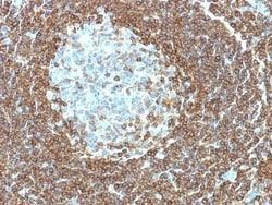
CD20 Antibody (109-3C2) - Azide and BSA Free, Novus Biologicals™
Manufacturer: Fischer Scientific
The price for this product is unavailable. Please request a quote
Antigen
CD20
Classification
Monoclonal
Concentration
1.0 mg/mL
Dilution
Flow Cytometry : 0.5 - 1 ug/million cells in 0.1 ml, Immunofluorescence : 0.5 - 1.0 ug/ml, CyTOF-ready
Gene Alias
B1, B-lymphocyte antigen CD20, B-lymphocyte cell-surface antigen B1, B-lymphocyte surface antigen B1, Bp35MGC3969, CD20 antigen, CD20 receptor, CD20S7, CVID5, LEU-16, Leukocyte surface antigen Leu-16, Membrane-spanning 4-domains subfamily A member 1, membrane-spanning 4-domains, subfamily A, member 1, MS4A2
Host Species
Mouse
Molecular Weight of Antigen
35 kDa
Quantity
0.2 mg
Research Discipline
Adaptive Immunity, B Cell Development and Differentiation Markers, Cancer Stem Cells, Cell Biology, Cytokine Research, Immunology, Signal Transduction, Stem Cell Markers, Tumor Biomarkers
Gene ID (Entrez)
931
Target Species
Human
Form
Purified
Applications
Flow Cytometry, Immunofluorescence, CyTOF
Clone
109-3C2
Conjugate
Unconjugated
Formulation
PBS with No Preservative
Gene Symbols
MS4A1
Immunogen
Stimulated human leukocytes
Purification Method
Protein A or G purified
Regulatory Status
RUO
Primary or Secondary
Primary
Test Specificity
Recognizes a protein of 30-33kDa, which is identified as CD20 (Workshop V; Code CD20.12. Workshop IV; Code B17). It recognizes an extracellular domain of CD20. It is a non-Ig differentiation antigen of B-cells and its expression is restricted to normal and neoplastic B-cells, being absent from all other leukocytes and tissues. CD20 is expressed by pre B-cells and persists during all stages of B-cell maturation but is lost upon terminal differentiation into plasma cells. The protein passes through the membrane 4 times with both ends in cytoplasm and exposes one short and one longer loop to the external environment. CD20 is not glycosylated in resting B-cells and its cytoplasmic domains are differentially phosphorylated upon activation. It acts as calcium channel involved in B cell activation and cell cycle progression.
Content And Storage
Store at 4C short term. Aliquot and store at -20C long term. Avoid freeze-thaw cycles.
Isotype
IgG3 κ
Related Products


Description
- CD20 Monoclonal specifically detects CD20 in Human samples
- It is validated for ELISA, Functional.
Compare Similar Items
Show Difference
Antigen: CD20
Classification: Monoclonal
Concentration: 1.0 mg/mL
Dilution: Flow Cytometry : 0.5 - 1 ug/million cells in 0.1 ml, Immunofluorescence : 0.5 - 1.0 ug/ml, CyTOF-ready
Gene Alias: B1, B-lymphocyte antigen CD20, B-lymphocyte cell-surface antigen B1, B-lymphocyte surface antigen B1, Bp35MGC3969, CD20 antigen, CD20 receptor, CD20S7, CVID5, LEU-16, Leukocyte surface antigen Leu-16, Membrane-spanning 4-domains subfamily A member 1, membrane-spanning 4-domains, subfamily A, member 1, MS4A2
Host Species: Mouse
Molecular Weight of Antigen: 35 kDa
Quantity: 0.2 mg
Research Discipline: Adaptive Immunity, B Cell Development and Differentiation Markers, Cancer Stem Cells, Cell Biology, Cytokine Research, Immunology, Signal Transduction, Stem Cell Markers, Tumor Biomarkers
Gene ID (Entrez): 931
Target Species: Human
Form: Purified
Applications: Flow Cytometry, Immunofluorescence, CyTOF
Clone: 109-3C2
Conjugate: Unconjugated
Formulation: PBS with No Preservative
Gene Symbols: MS4A1
Immunogen: Stimulated human leukocytes
Purification Method: Protein A or G purified
Regulatory Status: RUO
Primary or Secondary: Primary
Test Specificity: Recognizes a protein of 30-33kDa, which is identified as CD20 (Workshop V; Code CD20.12. Workshop IV; Code B17). It recognizes an extracellular domain of CD20. It is a non-Ig differentiation antigen of B-cells and its expression is restricted to normal and neoplastic B-cells, being absent from all other leukocytes and tissues. CD20 is expressed by pre B-cells and persists during all stages of B-cell maturation but is lost upon terminal differentiation into plasma cells. The protein passes through the membrane 4 times with both ends in cytoplasm and exposes one short and one longer loop to the external environment. CD20 is not glycosylated in resting B-cells and its cytoplasmic domains are differentially phosphorylated upon activation. It acts as calcium channel involved in B cell activation and cell cycle progression.
Content And Storage: Store at 4C short term. Aliquot and store at -20C long term. Avoid freeze-thaw cycles.
Isotype: IgG3 κ
Antigen: Laminin gamma 1
Classification: Monoclonal
Concentration: 1.0 mg/mL
Dilution: Flow Cytometry : 0.5 - 1 ug/million cells in 0.1 ml, Immunohistochemistry-Paraffin : 0.5 - 1.0 ug/ml, Immunofluorescence : 1 - 2 ug/ml, CyTOF-ready
Gene Alias: LAMB2Laminin-7 subunit gamma, Laminin B2 chain, laminin subunit gamma-1, laminin, gamma 1 (formerly LAMB2), Laminin-1 subunit gamma, Laminin-10 subunit gamma, Laminin-11 subunit gamma, Laminin-2 subunit gamma, Laminin-3 subunit gamma, Laminin-4 subunit gamma, Laminin-6 subunit gamma, Laminin-8 subunit gamma, Laminin-9 subunit gamma, MGC87297, S-LAM gamma, S-laminin subunit gamma
Host Species: Rat
Molecular Weight of Antigen: 210 kDa
Quantity: 0.1 mg
Research Discipline: Apoptosis, Cancer, Cytoskeleton Markers, Extracellular Matrix, Tumor Suppressors
Gene ID (Entrez): 3915
Target Species: Human, Mouse
Form: Purified
Applications: Flow Cytometry, Immunohistochemistry (Paraffin), Immunofluorescence, CyTOF
Clone: A5
Conjugate: Unconjugated
Formulation: PBS with No Preservative
Gene Symbols: LAMC1
Immunogen: Murine EHS laminin preparation
Purification Method: Protein A or G purified
Regulatory Status: RUO
Primary or Secondary: Primary
Test Specificity: Laminins are large hetero-trimeric, non-collagenous glycoproteins composed of alpha, beta, and gamma chains. This MAb reacts with laminin B2/1 chain of ∼210kDa and does not cross-react with other basement membrane components or fibronectin. Its specificity was established by immunoprecipitation and immunofluorescence of human skeletal muscle and kidney with laminin chain-specific MAbs. Epithelial sheets in vivo are separated from the mesenchymal elements of the stroma by a thin layer of a specialized type of extracellular matrix termed the basement membrane (BM). This structure consists of individual components, some of which are ubiquitous in BMs and some are not. The ubiquitous ones comprise laminin (LN), entactin/nidogen (EN), collagen type IV (CIV), and large heparan sulfate proteoglycan (HSPG), which interact specifically with each other to form a continuous and regular BM. Alterations of BM integrity, from local discontinuities up to complete loss, are described in many types of human and animal epithelial neoplasms. This MAb stains uniformly all human and murine basement membranes.
Content And Storage: Store at 4C short term. Aliquot and store at -20C long term. Avoid freeze-thaw cycles.
Isotype: IgG2a κ
Antigen: Laminin gamma 1
Classification: Monoclonal
Concentration: 1.0 mg/mL
Dilution: Flow Cytometry : 0.5 - 1 ug/million cells in 0.1 ml, Immunohistochemistry-Paraffin : 0.5 - 1.0 ug/ml, Immunofluorescence : 1 - 2 ug/ml, CyTOF-ready
Gene Alias: LAMB2Laminin-7 subunit gamma, Laminin B2 chain, laminin subunit gamma-1, laminin, gamma 1 (formerly LAMB2), Laminin-1 subunit gamma, Laminin-10 subunit gamma, Laminin-11 subunit gamma, Laminin-2 subunit gamma, Laminin-3 subunit gamma, Laminin-4 subunit gamma, Laminin-6 subunit gamma, Laminin-8 subunit gamma, Laminin-9 subunit gamma, MGC87297, S-LAM gamma, S-laminin subunit gamma
Host Species: Rat
Molecular Weight of Antigen: 210 kDa
Quantity: 0.2 mg
Research Discipline: Apoptosis, Cancer, Cytoskeleton Markers, Extracellular Matrix, Tumor Suppressors
Gene ID (Entrez): 3915
Target Species: Human, Mouse
Form: Purified
Applications: Flow Cytometry, Immunohistochemistry (Paraffin), Immunofluorescence, CyTOF
Clone: A5
Conjugate: Unconjugated
Formulation: PBS with No Preservative
Gene Symbols: LAMC1
Immunogen: Murine EHS laminin preparation
Purification Method: Protein A or G purified
Regulatory Status: RUO
Primary or Secondary: Primary
Test Specificity: Laminins are large hetero-trimeric, non-collagenous glycoproteins composed of alpha, beta, and gamma chains. This MAb reacts with laminin B2/1 chain of ∼210kDa and does not cross-react with other basement membrane components or fibronectin. Its specificity was established by immunoprecipitation and immunofluorescence of human skeletal muscle and kidney with laminin chain-specific MAbs. Epithelial sheets in vivo are separated from the mesenchymal elements of the stroma by a thin layer of a specialized type of extracellular matrix termed the basement membrane (BM). This structure consists of individual components, some of which are ubiquitous in BMs and some are not. The ubiquitous ones comprise laminin (LN), entactin/nidogen (EN), collagen type IV (CIV), and large heparan sulfate proteoglycan (HSPG), which interact specifically with each other to form a continuous and regular BM. Alterations of BM integrity, from local discontinuities up to complete loss, are described in many types of human and animal epithelial neoplasms. This MAb stains uniformly all human and murine basement membranes.
Content And Storage: Store at 4C short term. Aliquot and store at -20C long term. Avoid freeze-thaw cycles.
Isotype: IgG2a κ