CD43/Sialophorin Antibody (SPN/1094) - Azide and BSA Free, Novus Biologicals™
Manufacturer: Fischer Scientific
The price for this product is unavailable. Please request a quote
Antigen
CD43/Sialophorin
Classification
Monoclonal
Concentration
1.0 mg/mL
Dilution
Flow Cytometry : 0.5 - 1 ug/million cells in 0.1 ml, Immunohistochemistry-Paraffin : 0.5 - 1.0 ug/ml, Immunofluorescence : 1 - 2 ug/ml, CyTOF-ready
Gene Alias
CD43 antigen, CD43), Galactoglycoprotein, GALGP, Leukocyte sialoglycoprotein, Sialophorin, sialophorin (gpL115, leukosialin, CD43)
Host Species
Mouse
Purification Method
Protein A or G purified
Regulatory Status
RUO
Primary or Secondary
Primary
Test Specificity
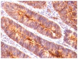
It recognizes a cell surface glycoprotein of 95/115/135kDa (depending upon the extent of glycosylation), identified as CD43. 70-90% of T-cell lymphomas and from 22-37% of B-cell lymphomas express CD43. No reactivity has been observed with reactive B-cells. So a B-lineage population that co-expresses CD43 is highly likely to be a malignant lymphoma, especially a low-grade lymphoma, rather than a reactive B-cell population. When CD43 antibody is used in combination with anti-CD20, effective immunophenotyping of the lymphomas in formalin-fixed tissues can be obtained. Co-staining of a lymphoid infiltrate with anti-CD20 and anti-CD43 argues against a reactive process and favors a diagnosis of lymphoma.
Content And Storage
Store at 4C short term. Aliquot and store at -20C long term. Avoid freeze-thaw cycles.
Isotype
IgG1 κ
Applications
Flow Cytometry, Immunohistochemistry (Paraffin), Immunofluorescence, CyTOF
Clone
SPN/1094
Conjugate
Unconjugated
Formulation
PBS with No Preservative
Gene Symbols
SPN
Immunogen
Recombinant human SPN protein
Quantity
0.2 mg
Research Discipline
B Cell Development and Differentiation Markers, Immunology
Gene ID (Entrez)
6693
Target Species
Human
Form
Purified
Related Products

Description
- CD43/Sialophorin Monoclonal specifically detects CD43/Sialophorin in Human samples
- It is validated for Western Blot, Flow Cytometry, Immunohistochemistry, Immunocytochemistry/Immunofluorescence, Immunohistochemistry-Paraffin, Immunofluorescence, CyTOF-ready.
Compare Similar Items
Show Difference
Antigen: CD43/Sialophorin
Classification: Monoclonal
Concentration: 1.0 mg/mL
Dilution: Flow Cytometry : 0.5 - 1 ug/million cells in 0.1 ml, Immunohistochemistry-Paraffin : 0.5 - 1.0 ug/ml, Immunofluorescence : 1 - 2 ug/ml, CyTOF-ready
Gene Alias: CD43 antigen, CD43), Galactoglycoprotein, GALGP, Leukocyte sialoglycoprotein, Sialophorin, sialophorin (gpL115, leukosialin, CD43)
Host Species: Mouse
Purification Method: Protein A or G purified
Regulatory Status: RUO
Primary or Secondary: Primary
Test Specificity: It recognizes a cell surface glycoprotein of 95/115/135kDa (depending upon the extent of glycosylation), identified as CD43. 70-90% of T-cell lymphomas and from 22-37% of B-cell lymphomas express CD43. No reactivity has been observed with reactive B-cells. So a B-lineage population that co-expresses CD43 is highly likely to be a malignant lymphoma, especially a low-grade lymphoma, rather than a reactive B-cell population. When CD43 antibody is used in combination with anti-CD20, effective immunophenotyping of the lymphomas in formalin-fixed tissues can be obtained. Co-staining of a lymphoid infiltrate with anti-CD20 and anti-CD43 argues against a reactive process and favors a diagnosis of lymphoma.
Content And Storage: Store at 4C short term. Aliquot and store at -20C long term. Avoid freeze-thaw cycles.
Isotype: IgG1 κ
Applications: Flow Cytometry, Immunohistochemistry (Paraffin), Immunofluorescence, CyTOF
Clone: SPN/1094
Conjugate: Unconjugated
Formulation: PBS with No Preservative
Gene Symbols: SPN
Immunogen: Recombinant human SPN protein
Quantity: 0.2 mg
Research Discipline: B Cell Development and Differentiation Markers, Immunology
Gene ID (Entrez): 6693
Target Species: Human
Form: Purified
Antigen: MUC-1
Classification: Monoclonal
Concentration: 1.0 mg/mL
Dilution: Flow Cytometry : 0.5 - 1 ug/million cells in 0.1 ml, Immunohistochemistry-Paraffin : 0.1 - 0.2 ug/ml, Immunofluorescence : 0.5 - 1.0 ug/ml, CyTOF-ready
Gene Alias: Breast carcinoma-associated antigen DF3, Carcinoma-associated mucin, CD227, CD227 antigen, DF3 antigen, EMA, episialin, H23 antigen, H23AG, KL-6, MAM6, MUC-1, MUC1/ZD, mucin 1, cell surface associated, mucin 1, transmembrane, mucin-1, Peanut-reactive urinary mucin, PEMMUC-1/SEC, PEMT, Polymorphic epithelial mucin, PUMMUC-1/X, tumor associated epithelial mucin, Tumor-associated epithelial membrane antigen, Tumor-associated mucin
Host Species: Mouse
Purification Method: Protein A or G purified
Regulatory Status: RUO
Primary or Secondary: Primary
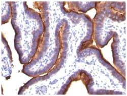
Test Specificity: In Western blotting, it recognizes proteins in MW range of 265-400kDa, identified as different glycoforms of EMA. The alpha subunit has cell adhesive properties. It can act both as an adhesion and an anti-adhesion protein. EMA may provide a protective layer on epithelial cells against bacterial and enzyme attack. The beta subunit contains a C-terminal domain, which is involved in cell signaling, through phosphorylations and protein-protein interactions. In immunohistochemical assays, it superbly stains routine formalin/paraffin carcinoma tissues. Antibody to EMA is useful as a pan-epithelial marker for detecting early metastatic loci of carcinoma in bone marrow or liver.
Content And Storage: Store at 4C short term. Aliquot and store at -20C long term. Avoid freeze-thaw cycles.
Isotype: IgG1 κ
Applications: Flow Cytometry, Immunohistochemistry (Paraffin), Immunofluorescence, CyTOF
Clone: MUC1/845
Conjugate: Unconjugated
Formulation: PBS with No Preservative
Gene Symbols: MUC1
Immunogen: Human milk-fat globule membranes (HMFGM)
Quantity: 0.1 mg
Research Discipline: Cancer, Cellular Markers, Extracellular Matrix
Gene ID (Entrez): 4582
Target Species: Human
Form: Purified
Antigen: MUC-1
Classification: Monoclonal
Concentration: 1.0 mg/mL
Dilution: Flow Cytometry : 0.5 - 1 ug/million cells in 0.1 ml, Immunohistochemistry-Paraffin : 0.1 - 0.2 ug/ml, Immunofluorescence : 0.5 - 1.0 ug/ml, CyTOF-ready
Gene Alias: Breast carcinoma-associated antigen DF3, Carcinoma-associated mucin, CD227, CD227 antigen, DF3 antigen, EMA, episialin, H23 antigen, H23AG, KL-6, MAM6, MUC-1, MUC1/ZD, mucin 1, cell surface associated, mucin 1, transmembrane, mucin-1, Peanut-reactive urinary mucin, PEMMUC-1/SEC, PEMT, Polymorphic epithelial mucin, PUMMUC-1/X, tumor associated epithelial mucin, Tumor-associated epithelial membrane antigen, Tumor-associated mucin
Host Species: Mouse
Purification Method: Protein A or G purified
Regulatory Status: RUO
Primary or Secondary: Primary
Test Specificity: In Western blotting, it recognizes proteins in MW range of 265-400kDa, identified as different glycoforms of EMA. The alpha subunit has cell adhesive properties. It can act both as an adhesion and an anti-adhesion protein. EMA may provide a protective layer on epithelial cells against bacterial and enzyme attack. The beta subunit contains a C-terminal domain, which is involved in cell signaling, through phosphorylations and protein-protein interactions. In immunohistochemical assays, it superbly stains routine formalin/paraffin carcinoma tissues. Antibody to EMA is useful as a pan-epithelial marker for detecting early metastatic loci of carcinoma in bone marrow or liver.
Content And Storage: Store at 4C short term. Aliquot and store at -20C long term. Avoid freeze-thaw cycles.
Isotype: IgG1 κ
Applications: Flow Cytometry, Immunohistochemistry (Paraffin), Immunofluorescence, CyTOF
Clone: MUC1/845
Conjugate: Unconjugated
Formulation: PBS with No Preservative
Gene Symbols: MUC1
Immunogen: Human milk-fat globule membranes (HMFGM)
Quantity: 0.2 mg
Research Discipline: Cancer, Cellular Markers, Extracellular Matrix
Gene ID (Entrez): 4582
Target Species: Human
Form: Purified