Tenascin C Antibody (SPM319) - Azide and BSA Free, Novus Biologicals™
Manufacturer: Fischer Scientific
The price for this product is unavailable. Please request a quote
Antigen
Tenascin C
Classification
Monoclonal
Concentration
1.0 mg/mL
Dilution
Flow Cytometry : 0.5 - 1 ug/million cells in 0.1 ml, Immunohistochemistry-Paraffin : 2 - 4 ug/ml, Immunofluorescence : 0.5 - 1.0 ug/ml, CyTOF-ready
Gene Alias
150-225, Cytotactin, Glioma-associated-extracellular matrix antigen, GMEM, GP 150-225, Hexabrachion, hexabrachion (tenascin C, cytotactin), hexabrachion (tenascin), HXBcytotactin, JI, MGC167029, Myotendinous antigen, neuronectin, tenascin, tenascin C, Tenascin-C, tenascin-C isoform 14/AD1/16, TN-C, TNGP
Host Species
Mouse
Purification Method
Protein A or G purified
Regulatory Status
RUO
Primary or Secondary
Primary
Test Specificity
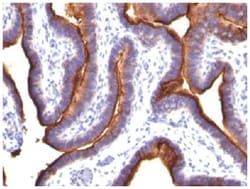
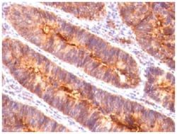
In Western blotting, it reacts with two bands of ∼MW of 210kDa and 300kDa, identified as two isoforms of Tenascin C. Specificity of this MAb is validated by sequential immunoprecipitation with a PAb against Tenascin C. Tenascin C is a multifunctional, disulfide-linked hexameric extracellular matrix glycoprotein expressed in association with mesenchymal epithelial interactions during development and in the neo-vasculature and stroma of undifferentiated tumors. In adults, it is restricted to certain epithelial-stromal interfaces and increases markedly in hyper-proliferative diseases and in stroma of many neoplasms, including gliomas, breast, squamous and lung carcinomas.
Content And Storage
Store at 4C short term. Aliquot and store at -20C long term. Avoid freeze-thaw cycles.
Isotype
IgG1 κ
Applications
Flow Cytometry, Immunohistochemistry (Paraffin), Immunofluorescence, CyTOF
Clone
SPM319
Conjugate
Unconjugated
Formulation
PBS with No Preservative
Gene Symbols
TNC
Immunogen
Human breast carcinoma
Quantity
0.2 mg
Research Discipline
Breast Cancer, Cancer, Extracellular Matrix, Neuroscience, Prostate Cancer
Gene ID (Entrez)
3371
Target Species
Human, Rat (Negative)
Form
Purified
Related Products

Description
- Tenascin C Monoclonal specifically detects Tenascin C in Human, Rat (Negative) samples
- It is validated for Flow Cytometry, Immunohistochemistry, Immunocytochemistry/Immunofluorescence, Immunohistochemistry-Paraffin, Immunofluorescence, CyTOF-ready.
Compare Similar Items
Show Difference
Antigen: Tenascin C
Classification: Monoclonal
Concentration: 1.0 mg/mL
Dilution: Flow Cytometry : 0.5 - 1 ug/million cells in 0.1 ml, Immunohistochemistry-Paraffin : 2 - 4 ug/ml, Immunofluorescence : 0.5 - 1.0 ug/ml, CyTOF-ready
Gene Alias: 150-225, Cytotactin, Glioma-associated-extracellular matrix antigen, GMEM, GP 150-225, Hexabrachion, hexabrachion (tenascin C, cytotactin), hexabrachion (tenascin), HXBcytotactin, JI, MGC167029, Myotendinous antigen, neuronectin, tenascin, tenascin C, Tenascin-C, tenascin-C isoform 14/AD1/16, TN-C, TNGP
Host Species: Mouse
Purification Method: Protein A or G purified
Regulatory Status: RUO
Primary or Secondary: Primary
Test Specificity: In Western blotting, it reacts with two bands of ∼MW of 210kDa and 300kDa, identified as two isoforms of Tenascin C. Specificity of this MAb is validated by sequential immunoprecipitation with a PAb against Tenascin C. Tenascin C is a multifunctional, disulfide-linked hexameric extracellular matrix glycoprotein expressed in association with mesenchymal epithelial interactions during development and in the neo-vasculature and stroma of undifferentiated tumors. In adults, it is restricted to certain epithelial-stromal interfaces and increases markedly in hyper-proliferative diseases and in stroma of many neoplasms, including gliomas, breast, squamous and lung carcinomas.
Content And Storage: Store at 4C short term. Aliquot and store at -20C long term. Avoid freeze-thaw cycles.
Isotype: IgG1 κ
Applications: Flow Cytometry, Immunohistochemistry (Paraffin), Immunofluorescence, CyTOF
Clone: SPM319
Conjugate: Unconjugated
Formulation: PBS with No Preservative
Gene Symbols: TNC
Immunogen: Human breast carcinoma
Quantity: 0.2 mg
Research Discipline: Breast Cancer, Cancer, Extracellular Matrix, Neuroscience, Prostate Cancer
Gene ID (Entrez): 3371
Target Species: Human, Rat (Negative)
Form: Purified
Antigen: Cyclin D1
Classification: Monoclonal
Concentration: 1.0 mg/mL
Dilution: Western Blot : 0.5 - 1.0 ug/ml, Flow Cytometry : 0.5 - 1 ug/million cells in 0.1 ml, Immunohistochemistry-Paraffin : 0.5 - 1.0 ug/ml, Immunofluorescence : 1 - 2 ug/ml, CyTOF-ready
Gene Alias: B-cell lymphoma 1 protein, BCL-1, BCL-1 oncogene, BCL1D11S287E, cyclin D1, cyclin D1 (PRAD1: parathyroid adenomatosis 1), G1/S-specific cyclin D1, G1/S-specific cyclin-D1, PRAD1 oncogene, PRAD1B-cell CLL/lymphoma 1, U21B31
Host Species: Mouse
Purification Method: Protein A or G purified
Regulatory Status: RUO
Primary or Secondary: Primary
Test Specificity: Recognizes a protein of 36kDa, identified as cyclin D1. Cyclin D1, one of the key cell cycle regulators, is a putative proto-oncogene overexpressed in a wide variety of human neoplasms. This antibody neutralizes the activity of cyclin D1 in vivo. About 60% of mantle cell lymphomas (MCL) contain a t(11; 14)(q13; q32) translocation resulting in over-expression of cyclin D1. This antibody is useful in identifying mantle cell lymphomas (cyclin D1 positive) from CLL/SLL and follicular lymphomas (cyclin D1 negative). Occasionally, hairy cell leukemia and plasma cell myeloma weakly express Cyclin D1.
Content And Storage: Store at 4C short term. Aliquot and store at -20C long term. Avoid freeze-thaw cycles.
Isotype: IgG2a κ
Applications: Western Blot, Flow Cytometry, Immunohistochemistry (Paraffin), Immunofluorescence, CyTOF
Clone: CCND1/809
Conjugate: Unconjugated
Formulation: PBS with No Preservative
Gene Symbols: CCND1
Immunogen: Recombinant human CCND1 protein
Quantity: 0.1 mg
Research Discipline: Cancer, Cell Cycle and Replication, Core ESC Like Genes, mTOR Pathway, Stem Cell Markers, Wnt Signaling Pathway
Gene ID (Entrez): 595
Target Species: Human
Form: Purified
Antigen: Cyclin D1
Classification: Monoclonal
Concentration: 1.0 mg/mL
Dilution: Western Blot : 0.5 - 1.0 ug/ml, Flow Cytometry : 0.5 - 1 ug/million cells in 0.1 ml, Immunohistochemistry-Paraffin : 0.5 - 1.0 ug/ml, Immunofluorescence : 1 - 2 ug/ml, CyTOF-ready
Gene Alias: B-cell lymphoma 1 protein, BCL-1, BCL-1 oncogene, BCL1D11S287E, cyclin D1, cyclin D1 (PRAD1: parathyroid adenomatosis 1), G1/S-specific cyclin D1, G1/S-specific cyclin-D1, PRAD1 oncogene, PRAD1B-cell CLL/lymphoma 1, U21B31
Host Species: Mouse
Purification Method: Protein A or G purified
Regulatory Status: RUO
Primary or Secondary: Primary
Test Specificity: Recognizes a protein of 36kDa, identified as cyclin D1. Cyclin D1, one of the key cell cycle regulators, is a putative proto-oncogene overexpressed in a wide variety of human neoplasms. This antibody neutralizes the activity of cyclin D1 in vivo. About 60% of mantle cell lymphomas (MCL) contain a t(11; 14)(q13; q32) translocation resulting in over-expression of cyclin D1. This antibody is useful in identifying mantle cell lymphomas (cyclin D1 positive) from CLL/SLL and follicular lymphomas (cyclin D1 negative). Occasionally, hairy cell leukemia and plasma cell myeloma weakly express Cyclin D1.
Content And Storage: Store at 4C short term. Aliquot and store at -20C long term. Avoid freeze-thaw cycles.
Isotype: IgG2a κ
Applications: Western Blot, Flow Cytometry, Immunohistochemistry (Paraffin), Immunofluorescence, CyTOF
Clone: CCND1/809
Conjugate: Unconjugated
Formulation: PBS with No Preservative
Gene Symbols: CCND1
Immunogen: Recombinant human CCND1 protein
Quantity: 0.2 mg
Research Discipline: Cancer, Cell Cycle and Replication, Core ESC Like Genes, mTOR Pathway, Stem Cell Markers, Wnt Signaling Pathway
Gene ID (Entrez): 595
Target Species: Human
Form: Purified