4420311
ENG Scientific Pneumocystis Carinii Stain Kit
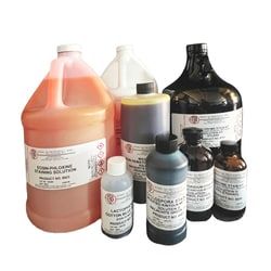
Modified Toluidine Blue O Stain allows the Pneumocystis carinii cysts to be visualized more easily after staining. For Pneumocystis Carinii Stain Kit (Modified Toluidine Blue O). Includes: 250 mL each of Solutions I, II, III and IV.
Manufacturer: Fischer Scientific
The price for this product is unavailable. Please request a quote
For Use With (Application)
In vitro diagnostic use
Related Products

Description
- Pneumocystis carinii is an opportunist which causes a diffuse interstitial pneumonia in patients with impaired immune systems
- The disease itself can be classified as epidemic or sporadic; the latter occurring in patients who have an underlying immunosuppression
- In this procedure, a sulfation reagent of glacial acetic acid and sulfuric acid (not included) is used for the removal of background material
- This allows the Pneumocystis carinii cysts to be visualized more easily after staining with Toluidine Blue 0
- Diagnosis of Pneumocystis carinii pneumonia can frequently be made on bronchoalveolar lavage (BAL) or on touch preparations of pulmonary tissue (open lung biopsies and transbronchial biopsies)
- Recommended Procedures: BAL Specimens: Using a 50 mL tube, centrifuge for 15 minutes at 2000 x g
- Aspirate all but the bottom 5 mL of supernatant
- With a Pasteur pipette, aspirate the sediment plus approximately 1 mL of the remaining 5 mL of fluid
- Transfer material to a 15 mL centrifuge tube, gently mix and prepare smear by spreading a drop over a 1 cmm area
- If concentrated specimen is very thick or mucoid, spread over entire slide with care being taken not to make the smear too thick
- Dry the slides on a heating block at 50 - 55°C
- Allow to cool before staining
- Touch Preparations: When dry, process in same manner as slides of BAL
- Preparation of Sulfation Reagent: In a dry coplin jar submerged in a plastic tub filled with cool water (not below 10°C), mix 45 mL of glacial acetic acid with 15 mL of concentrated sulfuric acid and stir gently with glass rod
- Carefully place smears in reagent for 10 minutes
- Mix immediately and again after 5 minutes
- Gently rinse with water for 10 minutes
- Drain excess water and stain with Sol
- I for 3 - 5 minutes
- Rinse with Sol
- II followed by Sol
- III for 10 seconds each
- Clear in Sol
- IV, two changes each for 10 seconds and mount
- Results: The cyst forms appear as lavender structures (cup-shaped) approximately 5 urn in diameter
- The cyst outline is distinct, and the internal region stains uniformly
- Note: A negative bronchoalveolar lavage does not rule out an infection with Pneumocystis carinii
- Other diagnostic procedures may be necessary.
Compare Similar Items
Show Difference
For Use With (Application): In vitro diagnostic use
For Use With (Application):
In vitro diagnostic use
For Use With (Application): __
For Use With (Application):
__
For Use With (Application): __
For Use With (Application):
__
For Use With (Application): __
For Use With (Application):
__