200009150
Invitrogen™ MitoTracker™ Dyes for Mitochondria Labeling
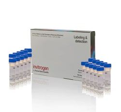
These mitochondria dyes are specially packaged in vials of 50 μg each to create freshly reconstituted dyes without the impact of freeze-thaw cycles to aid in a simplified sample prep for cell analysis workflow.
Manufacturer: Fischer Scientific
The price for this product is unavailable. Please request a quote
Content And Storage
Store in freezer -5°C to -30°C and protect from light.
For Use With (Equipment)
Fluorescence Microscope, Flow Cytometer, Microplate Reader
Detection Method
Fluorescence
Shipping Condition
Room Temperature
Product Type
Dye
Format
Special packaging
Quantity
20 x 50 μg
Product Line
MitoTracker™
Label Type
Fluorescent Dye
SubCellular Localization
Mitochondria/Organelle Stain
Related Products
Description
- Rosamine & carbocyanine-based staining dyes MitoTracker Orange CMTMRos, Red CMXRos, Orange CM-H2TMRos, Red CM-H2XRos, Red FM, Green FM, & Deep Red FM enable mitochondria visualization with fluorescent imaging
- These mitochondria dyes are specially packaged in vials of 50 μg each to create freshly reconstituted dyes without the impact of freeze-thaw cycles to aid in a simplified sample prep for cell analysis workflow
- Rosamine-based MitoTracker dyes for mitochondria labeling Cell-permeant MitoTracker probes stain active mitochondria in live cells for labeling and localization in fluorescent cell imaging
- Our rosamine-based mitochondrial staining dyes include MitoTracker Orange CMTMRos, a derivative of tetramethylrosamine, and MitoTracker Red CMXRos, a derivative of X-rosamine
- MitoTracker Orange CM-H2TMRos and MitoTracker Red CM-H2XRos are reduced, nonfluorescent versions of the rosamine-based MitoTracker dyes that fluoresce upon oxidation
- The fluorescent signal from the rosamine-based MitoTracker dyes is retained in the mitochondria even after aldehyde fixation and detergent permeabilization, making these mitochondria dyes flexible for many workflows including applications that require subsequent processing such as immunocytochemistry or in situ hybridization
- MitoTrackerRed and Orange are well suited for multicolor labeling experiments because their fluorescence is well resolved from the green fluorescence of other probes
- Carbocyanine-based MitoTracker dyes for mitochondria labeling Cell-permeant MitoTracker dyes stain active mitochondria in live cells for mitochondrial labeling and localization in fluorescent cell imaging
- The carbocyanine-based dyes MitoTracker Green FM and MitoTracker Red FM accumulate in active mitochondria but are not well-retained in mitochondria after aldehyde fixation
- The carbocyanine-based MitoTracker Deep Red FM stains active mitochondrial in live cells and is well-retained in mitochondria after aldehyde fixation and subsequent permeabilization with detergents for applications that require subsequent processing such as immunocytochemistry or in situ hybridization
- MitoTracker Red FM and MitoTracker Deep Red FM are well suited for multicolor labeling experiments because their red fluorescence is well resolved from the green fluorescence of other probes
- Benefits of using MitoTracker mitochondria labeling dyes MitoTracker dyes are provided in vials of 50 μg of lyophilized powder ready for reconstitution
- To label mitochondria, live cells are simply incubated with the MitoTracker probe of your choice
- The mitochondrial staining dyes passively diffuse across the plasma membrane and accumulate in active mitochondria
- The MitoTracker dyes are offered in a range of wavelengths and can be used for mitochondrial localization in multicolor experiments
- Conventional fluorescent stains such as tetramethylrosamine and rhodamine 123 are readily sequestered by active mitochondria and are reversible in dynamic membrane potential measurements as they easily wash out of cells upon loss in membrane potential
- In contrast, MitoTracker dyes contain a mildly thiol-reactive chloromethyl moiety so that mitochondrial staining is retained if the mitochondrial membrane potential is lost, allowing many of the MitoTracker dyes to be retained during cell fixation
- Throughout the cell life cycle, mitochondria use oxidizable substrates to produce an electrochemical proton gradient across the mitochondrial membrane (whose potential is negative), resulting in ATP production
- MitoTracker dyes are ideal probes for mitochondria staining in experiments studying the cell cycle or processes such as apoptosis and other end point assays
- MitoTracker dyes are also available for flow cytometry applications (Cat
- No
- M46750, M46751, M46752, and M46753)
- Related products for mitochondrial membrane potential For studying dynamic mitochondria membrane potential specifically, we recommend using JC-1 (cationic carbocyanine dye, Cat
- No
- T3168) or TMRM (tetramethyl rhodamine methyl ester, Cat
- No
- I34361) dyes
- The MitoProbe JC-1 Assay Kit (Cat
- No
- M34152) contains the JC-1 dye in addition to the potent mitochondrial membrane-potential disrupter, CCCP, which depolarizes the mitochondrial membrane
- These reagents can provide compensatory controls to correctly compensate green-to-red fluorescence ratio
- TMRM is used for the detection of dynamic measurement of mitochondrial membrane potential.
Compare Similar Items
Show Difference
Content And Storage: Store in freezer -5°C to -30°C and protect from light.
For Use With (Equipment): Fluorescence Microscope, Flow Cytometer, Microplate Reader
Detection Method: Fluorescence
Shipping Condition: Room Temperature
Product Type: Dye
Format: Special packaging
Quantity: 20 x 50 μg
Product Line: MitoTracker™
Label Type: Fluorescent Dye
SubCellular Localization: Mitochondria/Organelle Stain
Content And Storage:
Store in freezer -5°C to -30°C and protect from light.
For Use With (Equipment):
Fluorescence Microscope, Flow Cytometer, Microplate Reader
Detection Method:
Fluorescence
Shipping Condition:
Room Temperature
Product Type:
Dye
Format:
Special packaging
Quantity:
20 x 50 μg
Product Line:
MitoTracker™
Label Type:
Fluorescent Dye
SubCellular Localization:
Mitochondria/Organelle Stain


