AMEP4941
Celleste™ 6 Image Analysis Software, network license, one seat
Manufacturer: EVOS
Select a Size
| Pack Size | SKU | Availability | Price |
|---|---|---|---|
| Each of 1 | AMEP4941-Each-of-1 | In Stock | ₹ 7,80,530.00 |
AMEP4941 - Each of 1
In Stock
Quantity
1
Base Price: ₹ 7,80,530.00
GST (18%): ₹ 1,40,495.40
Total Price: ₹ 9,21,025.40
For Use With (Equipment)
EVOS
Related Products


Description
- Celleste 6 Image Analysis Software is a full-featured image analysis suite designed for biological applications
- Preconfigured analysis templates are optimized to deliver the most relevant data for a range of cell-based assays
- An icon-based wizard-driven workflow streamlines image analysis and delivers data faster
- A range of data display options allow users to view results across the plate as heat-maps, image montages, histograms, pie charts, and more
- Optional deconvolution, rendering, and GPU acceleration modules allow customization of the Celleste 6 capabilities to your cell models and analysis requirements
- Coupled with the powerful image acquisition capabilities of the EVOS imaging system, Celleste 6 software allows you to seamlessly capture, process, measure, analyze and share images and data
- This network license includes one seat that can be assigned by the administrator to multiple users across an internal network
- Only one user can access a seat at one time
- Additional seats are available (Cat
- No
- AMEP4869)
- Celleste 4 or Celleste 5 software can be upgraded to Celleste 6 software with either a single-user license upgrade (AMEP4870) or network license upgrade for one seat (AMPE4938)
- Celleste 6 Image Analysis Software offers these important advantages: • Icon-based wizard-driven workflow streamlines image analysis and eliminates guesswork • Preconfigured and optimized analysis templates for common applications help derive the most relevant results from image sets • Wide range of report formats to visualize results, create reports, and share images and data • Optional modules for 2D and 3D deconvolution, 3D visualization, and 3D analysis available to meet your specific needs Image analysis The Celleste 6 multi-channel analysis (MCA) tool is based on pre-configured algorithms and analysis templates that have been trained on representative data to optimally segment and classify images from a range of common cell-based assays
- Simply choose the app that corresponds to your assay of interest and follow the wizard-based workflow step by step from image to data generation
- Inspect images across the plate by clicking them or running the “play” function
- Then simply apply the analysis to the desired wells and immediately view results in a format most appropriate for the assay
- Choose from block diagram, graph, heat map, histogram, pie chart, scatter plot, or 3D
- Refine as desired and apply to the desired fields and wells in the data set
- Alternatively, use the full suite of powerful tools in Celleste 6 software to customize image analysis to your specific needs, including image adjustments, background correction, alignment and tiling, counting and sizing using powerful segmentation and classification tools, and measurements in up to four dimensions applied automatically over multiple channels, fields, and wells
- Display and export data with the same set of options as in the MCA workspace
- Measure and quantify Easily measure and analyze your images with a variety of measurement tools such as distance, region, angles, and area
- The ability to identify an object in time-lapse experiments can be used to track cell movement or migration
- It can also be used to track intensity changes over time, such as in cell death or target gene expression studies
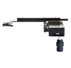
- This feature is particularly powerful in combination with the EVOS Onstage Incubator (Cat
- No
- AMC1000) that allows continuous monitoring of cells under controlled environmental conditions
- Report and share Upon completing the image analysis, a suite of annotation and reporting tools allow you to create presentation-ready images and data reports with a few clicks and to share them with others in PDF, PowerPoint™, and Excel™ format
- Save images as individual frames or movies according your needs
- Customize your toolset Choose from optional modules to customize Celleste 6 software to your cell models and analysis, visualization, and data generation needs
- Modules include 2D and 3D deconvolution, GPU acceleration, 3D rendering, visualization, and 3D analysis
- Celleste 6 software is a powerful image analysis solution that helps make your imaging research job easier and increases your productivity by seamlessly providing images and accurate data
- With the proven usability of EVOS imaging systems, you now have the tools that make it easy to capture, process, measure, analyze, and share your important images and data more effectively than ever.