17-10204
LentiBrite GFP-β-actin Lentiviral Biosensor
Manufacturer: Sigma Aldrich
Select a Size
| Pack Size | SKU | Availability | Price |
|---|---|---|---|
| 1 VIAL | 17-10204-1-VIAL | In Stock | ₹ 56,769.99 |
17-10204 - 1 VIAL
In Stock
Quantity
1
Base Price: ₹ 56,769.99
GST (18%): ₹ 10,218.598
Total Price: ₹ 66,988.588
manufacturer/tradename
Chemicon®LentiBrite
Quality Level
100
technique(s)
cell based assay: suitableimmunocytochemistry: suitableimmunofluorescence: suitablesingle cell analysis: suitabletransfection: suitable
UniProt accession no.
P60709
detection method
fluorometric
shipped in
dry ice
Related Products
Description
- General description: Actin is a highly abundant globular protein found in the cytosol of all eukaryotic cells that assembles in a head-to-tail arrangement to form double helical, polarized microfilaments. Actin microfilaments constitute one of the 3 major cytoskeletal structures, and they participate in many important cellular processes including muscle contraction, cell motility, cytokinesis, vesicle and organelle movement, cell signaling, the establishment and maintenance of cell junctions and cell shape. In vertebrates, three main groups of actin isoforms, alpha, beta, and gamma, have been identified; in most cell types, β-actin is the primary component of the cytoskeleton. Fusions of fluorescent proteins to β-actin have proven very useful tools in elucidating the dynamics of actin microfilaments in many cellular processes. EMD Millipore’s LentiBrite GFP-β-actin lentiviral particles provide bright fluorescence and precise localization to enable live cell analysis of actin microfilament dynamics in difficult-to-transfect cell types.
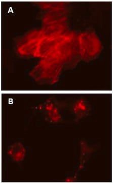
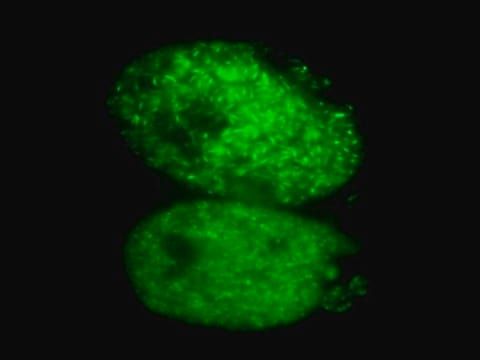
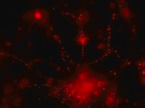
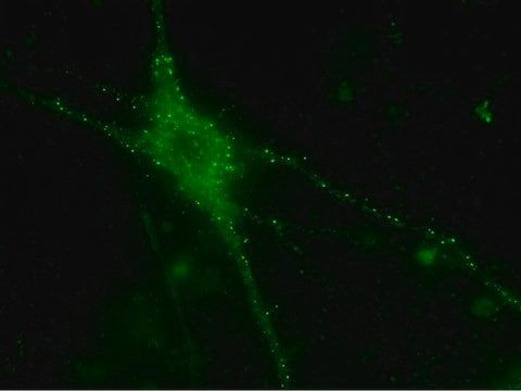
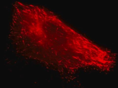
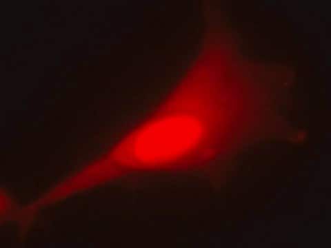
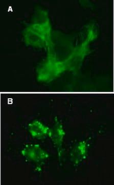
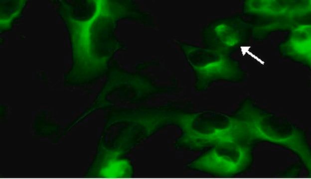
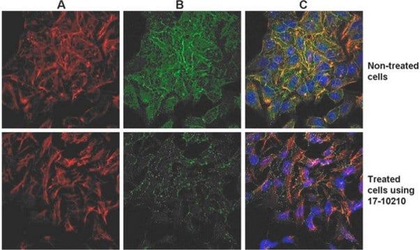
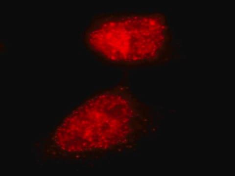
- Application: Immunocytochemistry comparison and inhibitor analysis:Similar to Figure 1 in the datasheet, HeLa cells were plated in a chamber slide and transduced with lentiviral particles at an MOI of 20 for 24 hours. After media replacement and 48 hours further incubation, cells were either untreated, incubated for 2 hours in complete media containing 2µM cytochalasin-D, or incubated for 2 hours in complete media containing 200nM jasplakinolide to induce actin depolymerization. Immunocytochemical staining (red) of the same fields of view with TRITC-conjugated Phalloidin reveals similar expression patterns to the GFP-protein (green). Hard-to-transfect cell types: Primary cell types HUVEC or HuMSC were plated in chamber slides and transduced with lentiviral particles at an MOI of 40 for 24 hours. Confocal microscopy imaging: HeLa cells were treated as in Figure 2A in the datasheet. Cells were then treated using ProteoExtract Native Cytoskeleton Enrichment Kit (Cat No. 17-10210). Unenriched & Enriched. Images were obtained by oil immersion confocal fluorescence microscopy. HT-1080 cells were treated as in Figures 2A and 2B in the datasheet. Confocal microscopy imaging: HT1080 cells were treated as in Figures 2A and 2B. For optimal fluorescent visualization, it is recommended to analyze the target expression level within 24-48 hrs after transfection/infection for optimal live cell analysis, as fluorescent intensity may dim over time, especially in difficult-to-transfect cell lines. Infected cells may be frozen down after successful transfection/infection and thawed in culture to retain positive fluorescent expression beyond 24-48 hrs. Length and intensity of fluorescent expression varies between cell lines. Higher MOIs may be required for difficult-to-transfect cell lines.
- Components: TagGFP2-β-actin Lentivirus: One vial containing 25 µL of lentiviral particles at a minimum of 3 x 10E8 infectious units (IFU) per mL. For lot-specific titer information, please see “Viral Titer” in the product specifications above. PromoterEF-1 (Elongation Factor-1)Multiplicty of Infection (MOI)MOI = Ratio of # of infectious lentiviral particles (IFU) to # of cells being infected.Typical MOI values for high transduction efficiency and signal intensity are in the range of 20-40. For this target, some cell types may require lower MOIs (e.g., HT-1080, human umbilical vein endothelial cells (HUVEC)), while others may require higher MOIs (e.g., human mesenchymal stem cells (HuMSC), HeLa, U2OS). NOTE: MOI should be titrated and optimized by the end user for each cell type and lentiviral target to achieve desired transduction efficiency and signal intensity.
- Quality: Evaluated by transduction of HT-1080 cells and fluorescent imaging performed for assessment of transduction efficiency.
- Physical form: PEG precipitation
- Storage and Stability: Storage and HandlingLentivirus is stable for at least 4 months from date of receipt when stored at -80°C. After first thaw, place immediately on ice and freeze in working aliquots at -80°C. Frozen aliquots may be stored for at least 2 months. Further freeze/thaws may result in decreased virus titer and transduction efficiency. IMPORTANT SAFETY NOTEReplication-defective lentiviral vectors, such as the 3rd Generation vector provided in this product, are not known to cause any diseases in humans or animals. However, lentiviruses can integrate into the host cell genome and thus pose some risk of insertional mutagenesis. Material is a Risk Group 2 and should be handled under BSL2 controls. A detailed discussion of biosafety of lentiviral vectors is provided in Pauwels, K. et al. (2009). State-of-the-art lentiviral vectors for research use: Risk assessment and biosafety recommendations. Curr. Gene Ther. 9: 459-474.
- Legal Information: CHEMICON is a registered trademark of Merck KGaA, Darmstadt, Germany
SAFETY INFORMATION
WGK
WGK 2
Compare Similar Items
Show Difference
manufacturer/tradename: Chemicon®LentiBrite
Quality Level: 100
technique(s): cell based assay: suitableimmunocytochemistry: suitableimmunofluorescence: suitablesingle cell analysis: suitabletransfection: suitable
UniProt accession no.: P60709
detection method: fluorometric
shipped in: dry ice
manufacturer/tradename:
Chemicon®LentiBrite
Quality Level:
100
technique(s):
cell based assay: suitableimmunocytochemistry: suitableimmunofluorescence: suitablesingle cell analysis: suitabletransfection: suitable
UniProt accession no.:
P60709
detection method:
fluorometric
shipped in:
dry ice
manufacturer/tradename: Chemicon®LentiBrite
Quality Level: 100
technique(s): cell based assay: suitableimmunocytochemistry: suitableimmunofluorescence: suitablesingle cell analysis: suitabletransfection: suitable
UniProt accession no.: P07437
detection method: fluorometric
shipped in: dry ice
manufacturer/tradename:
Chemicon®LentiBrite
Quality Level:
100
technique(s):
cell based assay: suitableimmunocytochemistry: suitableimmunofluorescence: suitablesingle cell analysis: suitabletransfection: suitable
UniProt accession no.:
P07437
detection method:
fluorometric
shipped in:
dry ice
manufacturer/tradename: Chemicon®ProteoExtract®
Quality Level: 100
technique(s): immunocytochemistry: suitableimmunofluorescence: suitable
UniProt accession no.: __
detection method: __
shipped in: dry ice
manufacturer/tradename:
Chemicon®ProteoExtract®
Quality Level:
100
technique(s):
immunocytochemistry: suitableimmunofluorescence: suitable
UniProt accession no.:
__
detection method:
__
shipped in:
dry ice
manufacturer/tradename: Chemicon®LentiBrite
Quality Level: 100
technique(s): cell based assay: suitableimmunocytochemistry: suitableimmunofluorescence: suitablesingle cell analysis: suitabletransfection: suitable
UniProt accession no.: Q06609
detection method: fluorometric
shipped in: dry ice
manufacturer/tradename:
Chemicon®LentiBrite
Quality Level:
100
technique(s):
cell based assay: suitableimmunocytochemistry: suitableimmunofluorescence: suitablesingle cell analysis: suitabletransfection: suitable
UniProt accession no.:
Q06609
detection method:
fluorometric
shipped in:
dry ice