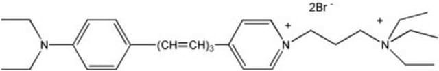
SCT127
BioTracker 640 Red C2(FM4-64) Synaptic Dye
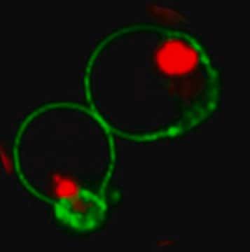
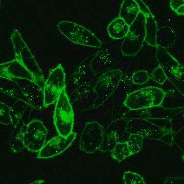
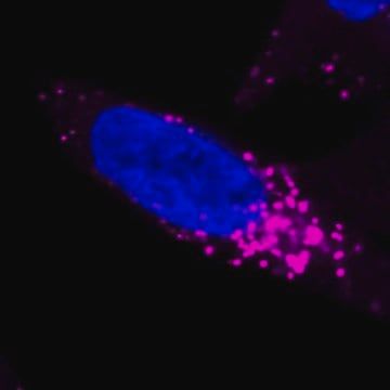
Live cell imaging synaptic dye that detects neural synaptic activitiy at neuromuscular junctions and synapses.
Manufacturer: Sigma Aldrich
Synonym(S): Live cell imaging probe
Select a Size
| Pack Size | SKU | Availability | Price |
|---|---|---|---|
| 5 MG | SCT127-5-MG | In Stock | ₹ 41,610.01 |
SCT127 - 5 MG
In Stock
Quantity
1
Base Price: ₹ 41,610.01
GST (18%): ₹ 7,489.802
Total Price: ₹ 49,099.812
technique(s)
cell based assay: suitable
detection method
fluorometric
shipped in
wet ice
Related Products


Description
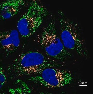
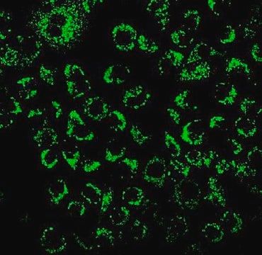
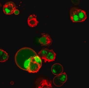
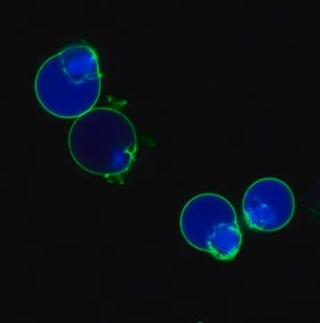
- General description: BioTracker synaptic dyes are a series of fluorescent cationic styryl dyes developed to follow synaptic activities at neuromuscular junctions or synapses. These dyes typically have a lipophilic tail at one end and a highly hydrophilic at the other end. In the presence of cells or tissue preparations, the dyes partition between the aqueous phase, where the dyes are virtually non-fluorescent, and the outer leaflet of the cell surface membranes, where the dyes insert the lipophilic end into the membranes and become intensely fluorescent. During endocytosis following nerve stimulation, the dyes become trapped inside the vesicles. Thus, after washing off the dyes on the cell surface, the fluorescent signal is proportional to the number of newly formed vesicles. On the other hand, during exocytosis, the dyes are released from the vesicles along with neurotransmitters, causing a decrease in fluorescent signal. As a result, the change in fluorescent intensity reflects the amount of endocytosis/exocytosis or synaptic activity.Spectral PropertiesAbsorbance (Membranes): 510 nmEmission (Membranes): 750 nm
- Application: Live cell fluorescent imaging
- Quality: Spectral PropertiesAbsorbance (Membranes): 510 nmEmission (Membranes): 750 nm
- Physical form: Lyophilized
- Storage and Stability: Store BioTracker 640 Red C2(FM4-64) Synaptic Dye at 2-8°C, protected from light. Stock solutions can be prepared at 10 mM and stored at 4°C or -20°C for six months or longer.
- Disclaimer: Unless otherwise stated in our catalog or other company documentation accompanying the product(s), our products are intended for research use only and are not to be used for any other purpose, which includes but is not limited to, unauthorized commercial uses, in vitro diagnostic uses, ex vivo or in vivo therapeutic uses or any type of consumption or application to humans or animals.
SAFETY INFORMATION
WGK
WGK 3
Flash Point(F)
Not applicable
Flash Point(C)
Not applicable
Compare Similar Items
Show Difference
technique(s): cell based assay: suitable
detection method: fluorometric
shipped in: wet ice
technique(s):
cell based assay: suitable
detection method:
fluorometric
shipped in:
wet ice
technique(s): cell based assay: suitable
detection method: fluorometric
shipped in: ambient
technique(s):
cell based assay: suitable
detection method:
fluorometric
shipped in:
ambient
technique(s): __
detection method: __
shipped in: __
technique(s):
__
detection method:
__
shipped in:
__
technique(s): __
detection method: __
shipped in: __
technique(s):
__
detection method:
__
shipped in:
__