BDB568263
Human T Cell Backbone Panel Kit, BD Horizon™
Manufacturer: BD Biosciences
Select a Size
| Pack Size | SKU | Availability | Price |
|---|---|---|---|
| Each of 1 | BDB568263-Each-of-1 | In Stock | ₹ 1,60,912.00 |
BDB568263 - Each of 1
In Stock
Quantity
1
Base Price: ₹ 1,60,912.00
GST (18%): ₹ 28,964.16
Total Price: ₹ 1,89,876.16
Applications
Flow Cytometry
Quantity
100 Tests
Content And Storage
Store undiluted at 4°C and protected from prolonged exposure to light. Do not freeze. The monoclonal antibody was purified from tissue culture supernatant or ascites by affinity chromatography. The antibody was conjugated to the dye under optimum conditions and unconjugated antibody and free dye were removed. The antibody was conjugated to the dye under optimum conditions and unreacted dye was removed.
Conjugate
Fluorochromes (BV786, R718, PE-Cy7, BV510, BV711)
Target Species
Human
Description
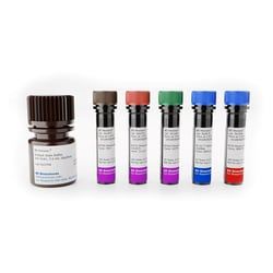
- The Human T Cell Backbone Panel contains five individual vials of fluorochrome-conjugated antibodies against T cell core markers [CD3, CD4, CD8, CD45RA, CD197 (CCR7)] conventionally used to identify different maturational states of CD4+ and CD8+ T cells (naïve, central memory, effector memory, effector memory RA)
- The kit also contains the BD HorizonTM Brilliant Stain Buffer for optimal performance
- The Human T Cell Backbone Panel is strategically designed to be complemented with 4-5 drop-in fluorochromes, ie, fluorescent reagents specific for biomarkers of choice, depending on instrument configuration, with minimal panel design effort
- Assignment of fluorochromes for drop-ins have minimal impact on the resolution of the backbone panel
- The fluorochromes used for the backbone panel have minimal resolution impact on the detectors allocated for drop-in fluorochromes and the specified fluorochromes available for drop-in do not impact each other
- The Human T Cell Backbone Panel is compatible with intracellular stain and transcription factor analysis.
Compare Similar Items
Show Difference
Applications: Flow Cytometry
Quantity: 100 Tests
Content And Storage: Store undiluted at 4°C and protected from prolonged exposure to light. Do not freeze. The monoclonal antibody was purified from tissue culture supernatant or ascites by affinity chromatography. The antibody was conjugated to the dye under optimum conditions and unconjugated antibody and free dye were removed. The antibody was conjugated to the dye under optimum conditions and unreacted dye was removed.
Conjugate: Fluorochromes (BV786, R718, PE-Cy7, BV510, BV711)
Target Species: Human
Applications:
Flow Cytometry
Quantity:
100 Tests
Content And Storage:
Store undiluted at 4°C and protected from prolonged exposure to light. Do not freeze. The monoclonal antibody was purified from tissue culture supernatant or ascites by affinity chromatography. The antibody was conjugated to the dye under optimum conditions and unconjugated antibody and free dye were removed. The antibody was conjugated to the dye under optimum conditions and unreacted dye was removed.
Conjugate:
Fluorochromes (BV786, R718, PE-Cy7, BV510, BV711)
Target Species:
Human
Applications: Flow Cytometry
Quantity: 50 μg
Content And Storage: Store undiluted at 4°C and protected from prolonged exposure to light. Do not freeze. The monoclonal antibody was purified from tissue culture supernatant or ascites by affinity chromatography. The antibody was conjugated to the dye under optimum conditions and unconjugated antibody and free dye were removed.
Conjugate: Brilliant Violet 786
Target Species: Human
Applications:
Flow Cytometry
Quantity:
50 μg
Content And Storage:
Store undiluted at 4°C and protected from prolonged exposure to light. Do not freeze. The monoclonal antibody was purified from tissue culture supernatant or ascites by affinity chromatography. The antibody was conjugated to the dye under optimum conditions and unconjugated antibody and free dye were removed.
Conjugate:
Brilliant Violet 786
Target Species:
Human
Applications: Flow Cytometry
Quantity: 0.1 mg
Content And Storage: Store undiluted at 4°C and protected from prolonged exposure to light. Do not freeze. The monoclonal antibody was purified from tissue culture supernatant or ascites by affinity chromatography. The antibody was conjugated to the dye under optimum conditions and unconjugated antibody and free dye were removed.
Conjugate: PE
Target Species: Human
Applications:
Flow Cytometry
Quantity:
0.1 mg
Content And Storage:
Store undiluted at 4°C and protected from prolonged exposure to light. Do not freeze. The monoclonal antibody was purified from tissue culture supernatant or ascites by affinity chromatography. The antibody was conjugated to the dye under optimum conditions and unconjugated antibody and free dye were removed.
Conjugate:
PE
Target Species:
Human
Applications: Flow Cytometry
Quantity: 25 μg
Content And Storage: Store undiluted at 4°C and protected from prolonged exposure to light. Do not freeze. The monoclonal antibody was purified from tissue culture supernatant or ascites by affinity chromatography. The antibody was conjugated to the dye under optimum conditions and unconjugated antibody and free dye were removed.
Conjugate: PE
Target Species: Human
Applications:
Flow Cytometry
Quantity:
25 μg
Content And Storage:
Store undiluted at 4°C and protected from prolonged exposure to light. Do not freeze. The monoclonal antibody was purified from tissue culture supernatant or ascites by affinity chromatography. The antibody was conjugated to the dye under optimum conditions and unconjugated antibody and free dye were removed.
Conjugate:
PE
Target Species:
Human
